

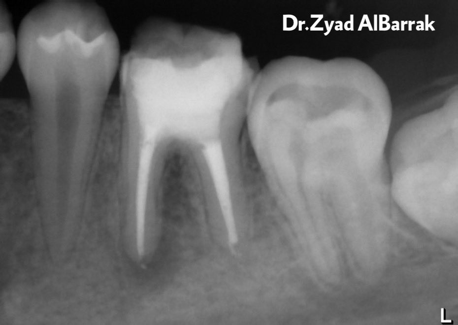
22 yrs old pt, saudi female, medically fit, She’s referred from undergrade student to re-treat #36.
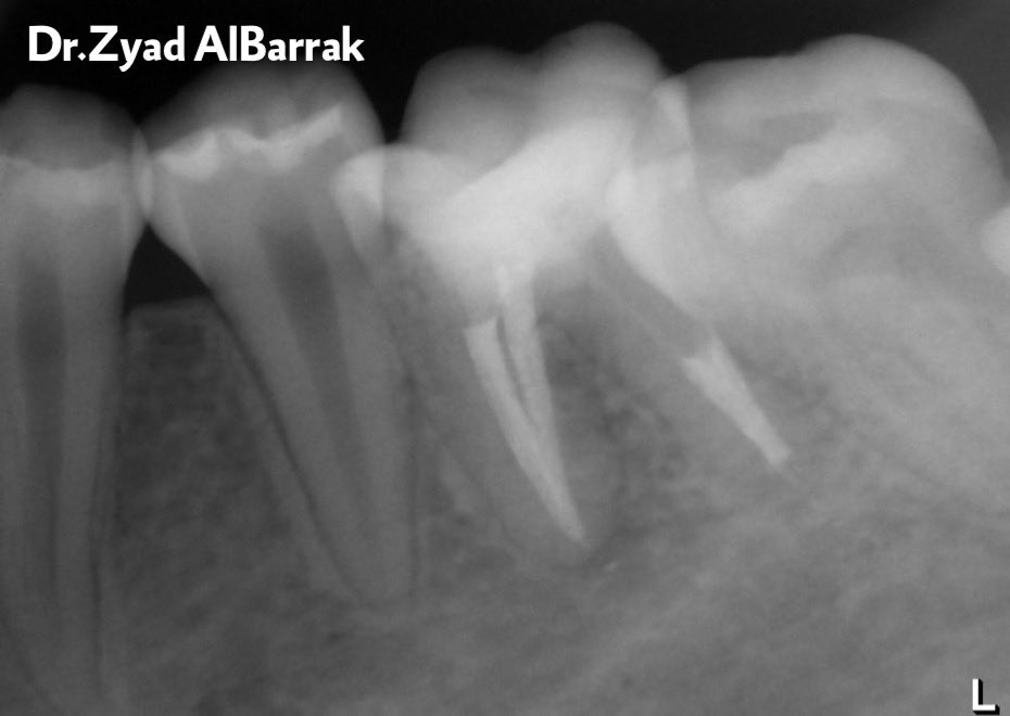
After radiograph interpretation:
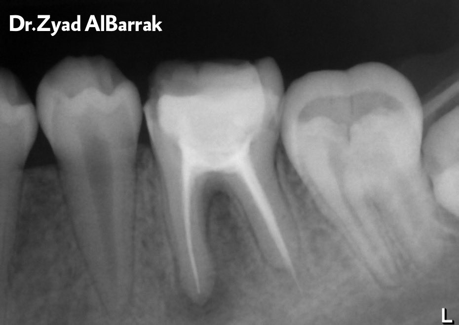
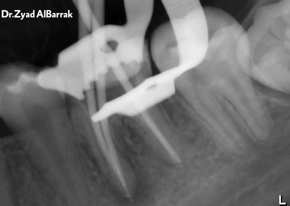
#36 > endo-treated tooth with apical radiolucency + broken lamina dura and extruded gutta percha on distal canal.
After radiograph interpretation:
#36 > endo-treated tooth with apical radiolucency + broken lamina dura and extruded gutta percha on distal canal.
Clinical Examination:
#36 endo-treated
#36 RCT was did before 3 years ago
#36 pain on percussion and palpation.
Pocket depth 2 mm all around.
Dignosis:
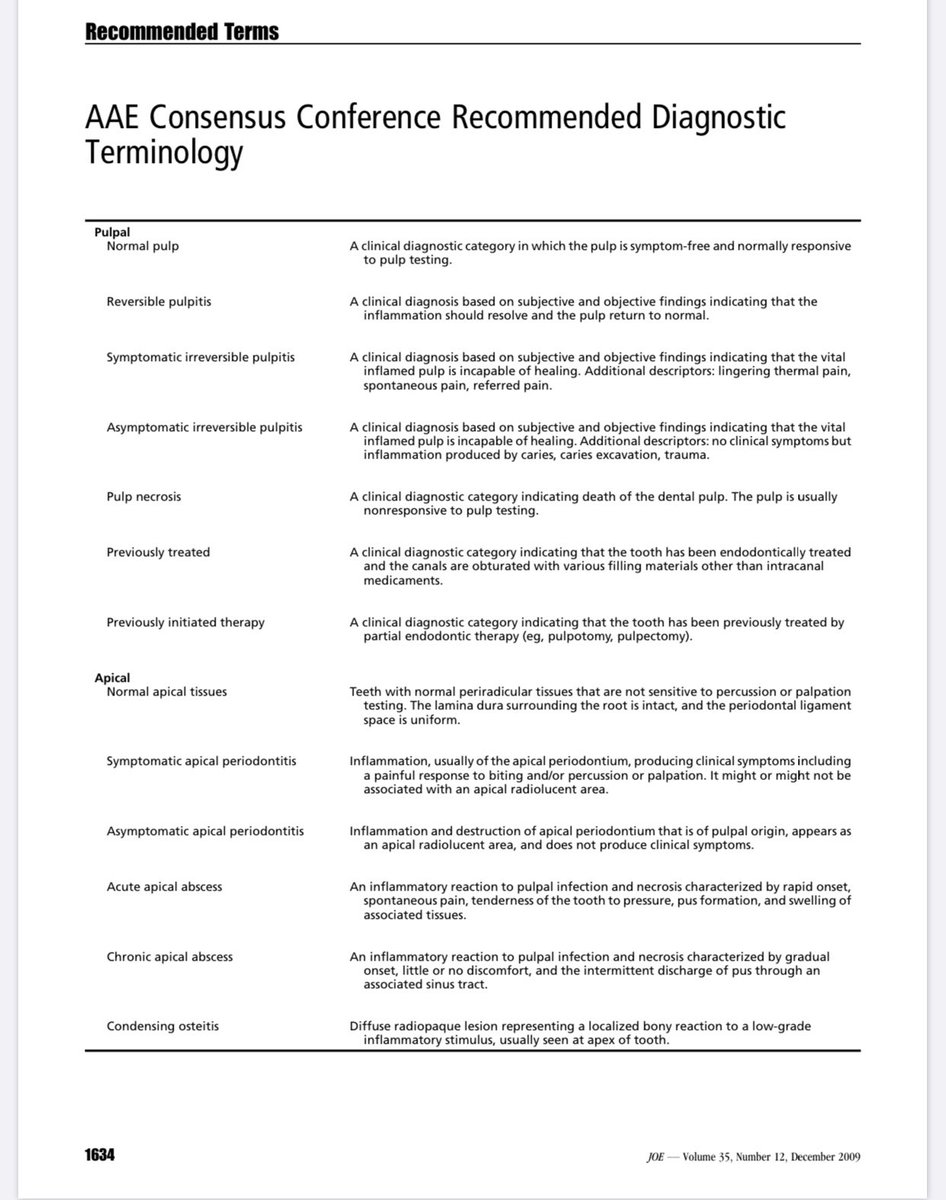
Previously treated with Symptomatic apical periodontitis.
#36 endo-treated
#36 RCT was did before 3 years ago
#36 pain on percussion and palpation.
Pocket depth 2 mm all around.
Dignosis:
Previously treated with Symptomatic apical periodontitis.
Tx plan:
1- Non-Surgical Re-Treatment
2- If the lesion not healed or decrease the size during follow up, will go for Surgical Tx.
1- Non-Surgical Re-Treatment
2- If the lesion not healed or decrease the size during follow up, will go for Surgical Tx.
Causes of failure of initial endodontic therapy:
➢Poor cleaning and obturation
➢Coronal leakage
➢Untreated canals
➢Poor access cavity
➢Complications of instrumentation
➢Overextension of root canal filling
➢Persistent intracanal or extracanal infection
➢Radicular cyst
➢Poor cleaning and obturation
➢Coronal leakage
➢Untreated canals
➢Poor access cavity
➢Complications of instrumentation
➢Overextension of root canal filling
➢Persistent intracanal or extracanal infection
➢Radicular cyst
Tx procedure:
1st visit:
Remove all GP by profile ,(04- 30) file and chloroform to get access to all the canals.
Irrigate with NaOCL 5.25%, saline, CHX.
1st visit:
Remove all GP by profile ,(04- 30) file and chloroform to get access to all the canals.
Irrigate with NaOCL 5.25%, saline, CHX.
According to Ferreira, 2001
In comparison between K-Flexofiles with chloroform, Hedstrom files with chloroform, ProFiles 0.04 with chloroform, ProFiles 0.04 alone in removing the gutta-percha from obturated root canals :
In comparison between K-Flexofiles with chloroform, Hedstrom files with chloroform, ProFiles 0.04 with chloroform, ProFiles 0.04 alone in removing the gutta-percha from obturated root canals :
-There was no statistically significant difference in canal cleanliness between K-Flexofiles and ProFiles.
-ProFiles were significantly faster than hand files.
-ProFiles were significantly faster than hand files.
According to Ray,1995
Good coronal restorations and endodontic treatment resulted in the absence of periradicular inflammation in91.4%,whereas poor coronal restorations and endodontic treatment resulted in the presence of periradicular inflammation in 81.9%of the teeth examined.
Good coronal restorations and endodontic treatment resulted in the absence of periradicular inflammation in91.4%,whereas poor coronal restorations and endodontic treatment resulted in the presence of periradicular inflammation in 81.9%of the teeth examined.
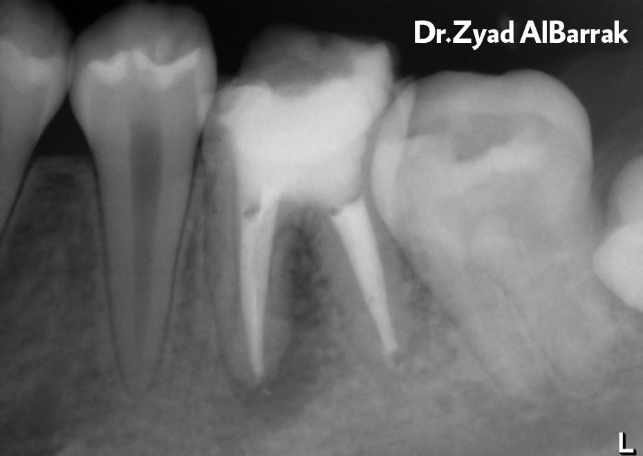
WLs determined by EAL and confirmed radiographically.
All canals cleaned & shaped by Protaper Next rotary files and 5.25% NaOCl
According to Vertucci FJ,1984 regarding configuration of root canals
All canals cleaned & shaped by Protaper Next rotary files and 5.25% NaOCl
According to Vertucci FJ,1984 regarding configuration of root canals
Master cone and obturation was done by hybrid technique ( lateral and vertical compaction )
According to Ng YL, 2008
1- The pooled estimated success rate of secondary root canal treatment was 77%.
2- The presence of pre-operative periapical lesion, apical extent of root filling and quality of coronal restoration proved significant prognostic factors.
1- The pooled estimated success rate of secondary root canal treatment was 77%.
2- The presence of pre-operative periapical lesion, apical extent of root filling and quality of coronal restoration proved significant prognostic factors.
3-The outcome of 2 RCT should therefore be similar to 1 RCT as long as access to the apical infection can be re-established
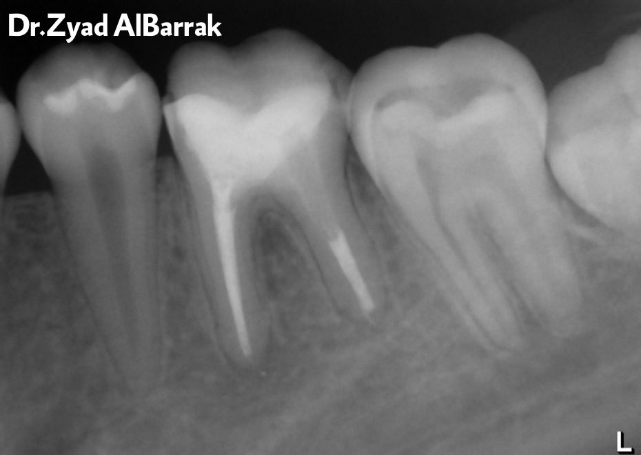
1 year follow up:
No signs and symptom in follow up appointment.
Healing in process.
No signs and symptom in follow up appointment.
Healing in process.
Loading suggestions...