

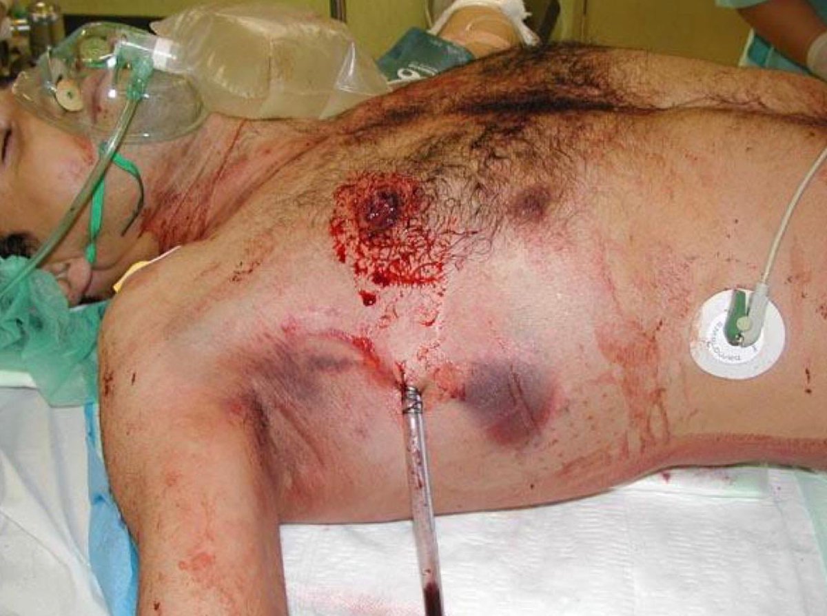
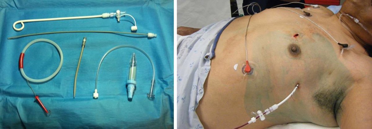
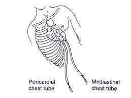
❇️ A chest tube (chest drain): is a flexible plastic tube that is inserted through the chest (lateral / mediastinal) into the pleural space.
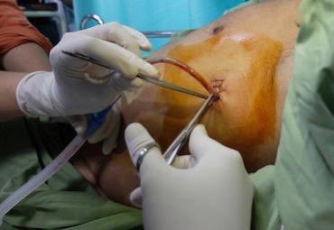
-The insertion of a chest tube is a medical procedure, performed by a physician.
-The insertion of a chest tube is a medical procedure, performed by a physician.
❇️It is used to:
-Remove excess air or fluid, or pus from the intrathoracic space.
-To help regain negative pressure.
-To assist in the re-expansion of remaining lung tissue.
-Chest tube used during and immediately after thoracic surgery (e.g. open heart surgery) mediastinal.
-Remove excess air or fluid, or pus from the intrathoracic space.
-To help regain negative pressure.
-To assist in the re-expansion of remaining lung tissue.
-Chest tube used during and immediately after thoracic surgery (e.g. open heart surgery) mediastinal.
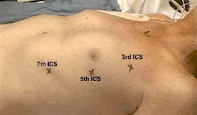
❇️Sites of tube insertion:
*Remember*
Air rises and fluid falls location depends on what is being drained
1. Free air in the pleural space (pneumothorax):
Tube is placed above the 2nd to 3rd intercostal space midclavicular or anterior axillary line.
*Remember*
Air rises and fluid falls location depends on what is being drained
1. Free air in the pleural space (pneumothorax):
Tube is placed above the 2nd to 3rd intercostal space midclavicular or anterior axillary line.
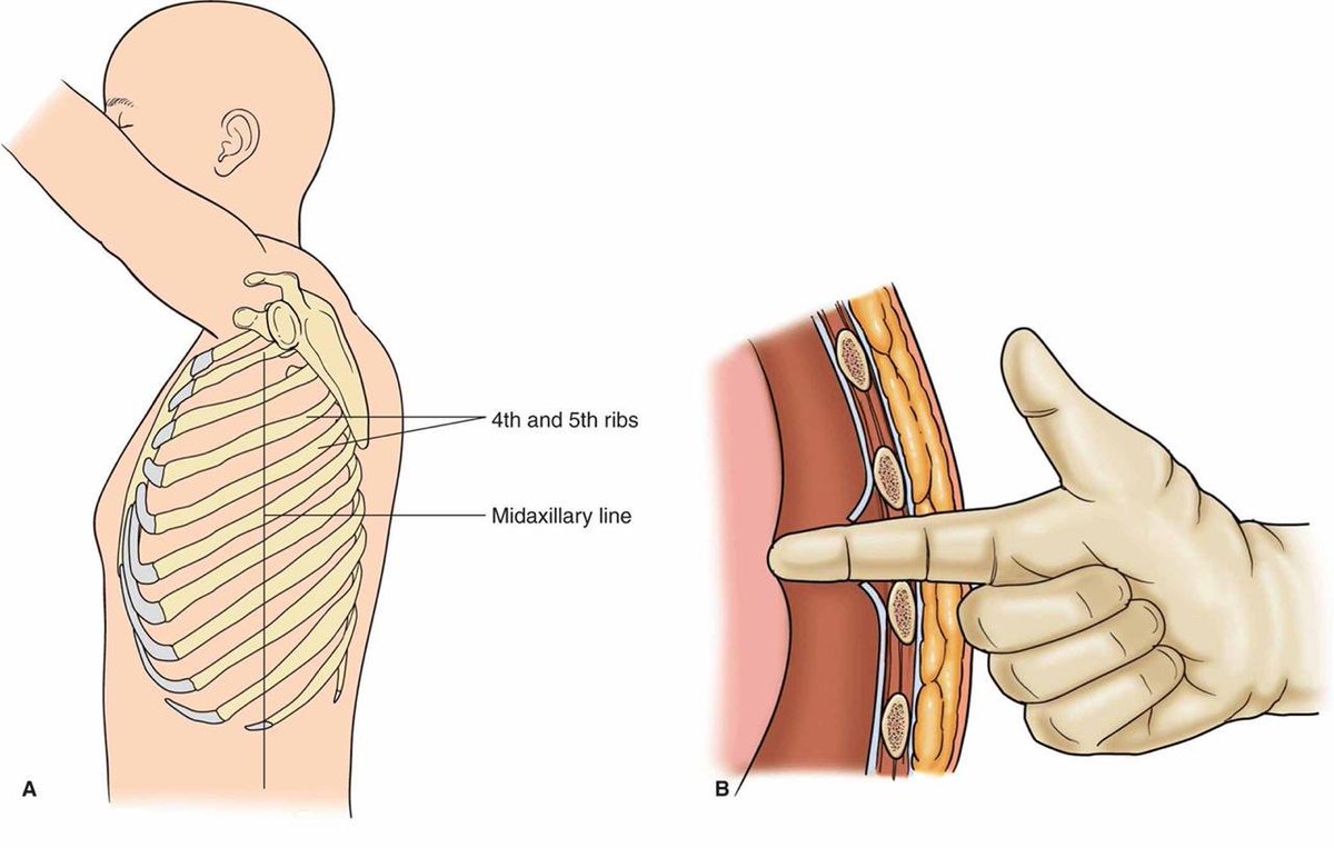
2. Fluids (Hemothorax, pleural effusion): gravitate to the most dependent point, tube is placed at the 4th to 5th, 6th to 7th intercostal space-midaxillary line.
3. Mediastinal tubes: are put in place after cardiac surgery to drain fluid from around heart.
3. Mediastinal tubes: are put in place after cardiac surgery to drain fluid from around heart.
❇️ Nursing considerations :
-Assess cardiopulmonary statues of the patient and vitals every 2 hrs.
-Verify that all connection tube are patent.
-Monitor characteristics of drainage fluid in colour, amount, consistency and marked significant increase or decrease output.
-Assess cardiopulmonary statues of the patient and vitals every 2 hrs.
-Verify that all connection tube are patent.
-Monitor characteristics of drainage fluid in colour, amount, consistency and marked significant increase or decrease output.
-Keep system below the patient chest level.
-Observe the tube for any kink or clot lead to obstructions.
-Monitor fluctuations (tidaling) and volume at least q shift.
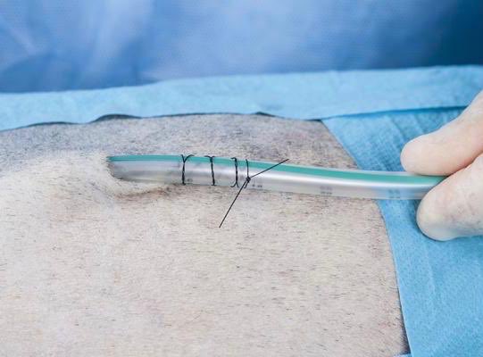
-Assess actual chest tube insertion site for signs of infection.
-Observe the tube for any kink or clot lead to obstructions.
-Monitor fluctuations (tidaling) and volume at least q shift.
-Assess actual chest tube insertion site for signs of infection.
-Change dressing every other day or when ordered.
-Assess the suction control chamber for air bubbling & at level ordered.
-Bubbling in the suction control chamber indicates the system is functioning properly.
-Assess the suction control chamber for air bubbling & at level ordered.
-Bubbling in the suction control chamber indicates the system is functioning properly.
-Monitor air leak: Water seal chamber is a window into the pleural space. If air is leaving the chest, bubbling will be seen here.
❖ Bubbling in water seal chamber may be present with pneumothorax. if worsens (continuous) or occurs in absence of pneumothorax, this may indicate air leak.
*keep remembering* that continuous bubbling in this chamber indicates air leakage.
*keep remembering* that continuous bubbling in this chamber indicates air leakage.
❇️More informations about bubbling, chest tubes and drainage systems 👇🏻
youtu.be
❇️Clamping:
You will only clamp for the following reasons:
-Prior to removing chest tube to determine if patient can do without chest tube(s).
youtu.be
❇️Clamping:
You will only clamp for the following reasons:
-Prior to removing chest tube to determine if patient can do without chest tube(s).
-Assessing for air leak (clamp only briefly)
-Changing the chest drainage unit (clamp only briefly).
-Performing physician-ordered procedure.
-Some instances when sudden large volumes of fluid are evacuated.
-Changing the chest drainage unit (clamp only briefly).
-Performing physician-ordered procedure.
-Some instances when sudden large volumes of fluid are evacuated.
❇️Air leak:How to detect
Clamp the chest tube momentarily, beginning at the patient.Look at the chamber to see whether the bubbling has stopped.
1.If you clamp and the bubbling goes away,the leak is coming from the chest
Action:reinforce dressing with Jelonet,inform physician
Clamp the chest tube momentarily, beginning at the patient.Look at the chamber to see whether the bubbling has stopped.
1.If you clamp and the bubbling goes away,the leak is coming from the chest
Action:reinforce dressing with Jelonet,inform physician
2. If you clamp at the chest and the bubbling persists, the leak is between the clamp and the water seal chamber.
Action: change the drainage system.
Action: change the drainage system.
❇️ Complications:
-Lung laceration (if placed too deep)
-Diaphragm / Abdominal cavity penetration (if placed too low)
-Tube placed subcutaneously (not in thoracic cavity)
-Tube placed too far (causes pain)
-Tube falls out (if not secured)
-Blocked tube (clot)
-Lung laceration (if placed too deep)
-Diaphragm / Abdominal cavity penetration (if placed too low)
-Tube placed subcutaneously (not in thoracic cavity)
-Tube placed too far (causes pain)
-Tube falls out (if not secured)
-Blocked tube (clot)
-Empyema (collection of pus within a naturally existing anatomical cavity, such as the lung pleura) causes infection.
❇️Removal indications:
1. One day after cessation of air leak.
2. Drainage less than 50-100ml of fluid per day.
3. 1-3 days post cardiac surgery.
4. 2-6 days post thoracic surgery.
5. Hemoserus drainage from around the chest tube insertion site.
1. One day after cessation of air leak.
2. Drainage less than 50-100ml of fluid per day.
3. 1-3 days post cardiac surgery.
4. 2-6 days post thoracic surgery.
5. Hemoserus drainage from around the chest tube insertion site.
Done by: @Perfectionology
Loading suggestions...