

Severe pain in the left ankle after total left hip replacement. Differential diagnosis?


CRPS- Complex regional pain syndrome. Not an imaging diagnosis. The clinical context is important. Diffuse persistent pain with Vasomotor disturbances, Trophic changes and Limited range of motion or immobility of joints.
CRPS type 1: No detectable nerve lesion- Replaces term reflex sympathetic dystrophy (RSD)
CRPS type 2: Detectable nerve lesion with resultant pain along distribution of nerve- Replaces term causalgia.
This case is type 2- as it is post THR with possible nerve injury
CRPS type 2: Detectable nerve lesion with resultant pain along distribution of nerve- Replaces term causalgia.
This case is type 2- as it is post THR with possible nerve injury
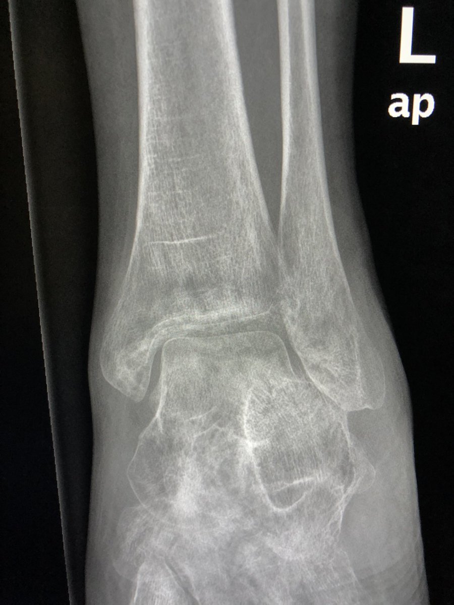
Radiographs may show diffuse regional osteoporosis- as in our case. Subcortical changes may predominate- as in our case (rounded areas of demineralisation disproportionate to the diffuse osteopenia). Trophic changes in soft tissues may be seen.
In the absence of typical clinical syndrome, these findings just represents osteopenia, often disuse.
This is one of those MSK conditions where bone scan does have a role and possible prognostic value. High intensity on bone scan predictive of good response to early therapy.
In severe osteopenia, you will see cortical tunnelling, which is seen in this case!
Talus may start looking like positive Hawkins sign. In fact, one of my residents raised the concern for talus AVN- due to subchondral linear osteopenia, which looks like positive Hawkins sign.
Loading suggestions...