

Essential of lung/diaphragm #POCUS tutorial 🧵
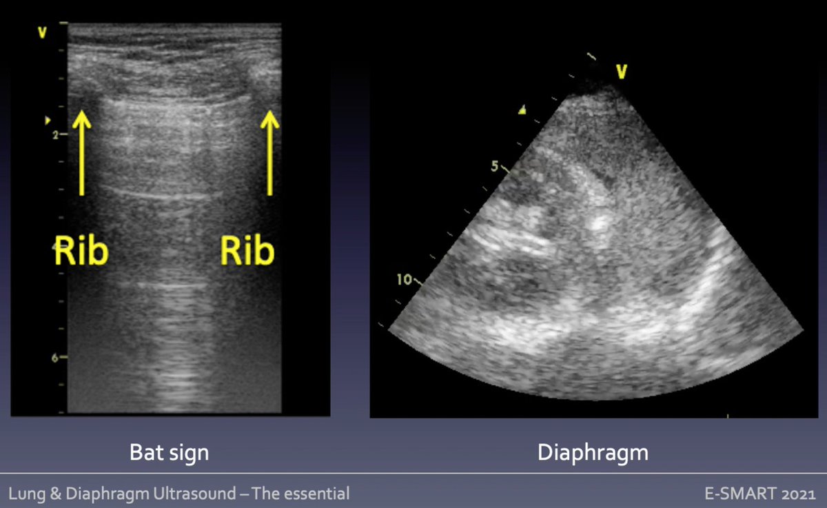
🦇 sign identify lung in ICS (ribs acoustic shadows)
__ pleural line moves w ventilation: visceral/parietal pleura sliding
🅰️ lines horizontal reverberation artifacts similar to pleural line (repeats = distance)
Mongodi S #eSMART2021
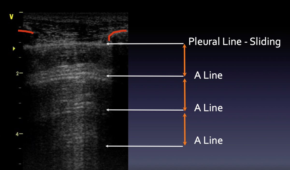
🦇 sign identify lung in ICS (ribs acoustic shadows)
__ pleural line moves w ventilation: visceral/parietal pleura sliding
🅰️ lines horizontal reverberation artifacts similar to pleural line (repeats = distance)
Mongodi S #eSMART2021
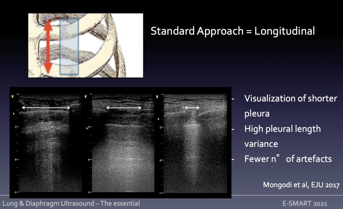
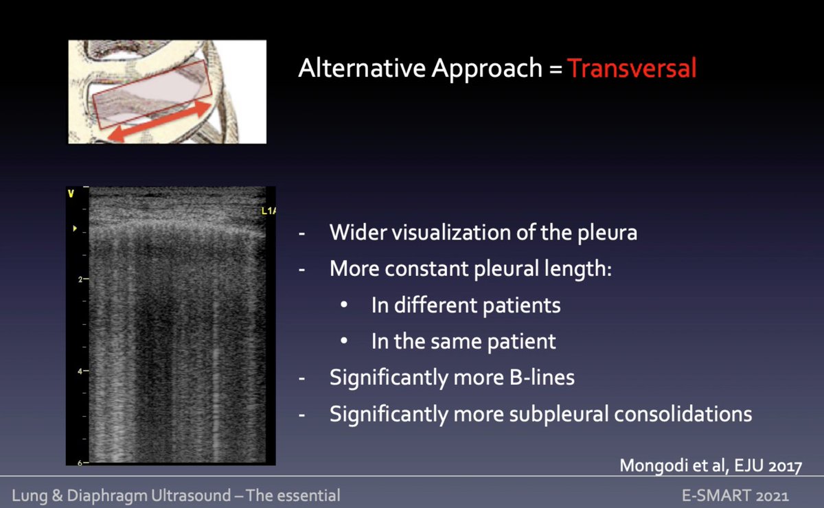
Approach for lung #ultrasound
➡️longitudinal (standard) = transversal to ICS; smaller ICS = smaller window
➡️transversal (alternative) = aligned to ICS
Check
📰 bit.ly
📰 bit.ly
🧵Mongodi S tutorial #eSMART2021 #POCUS
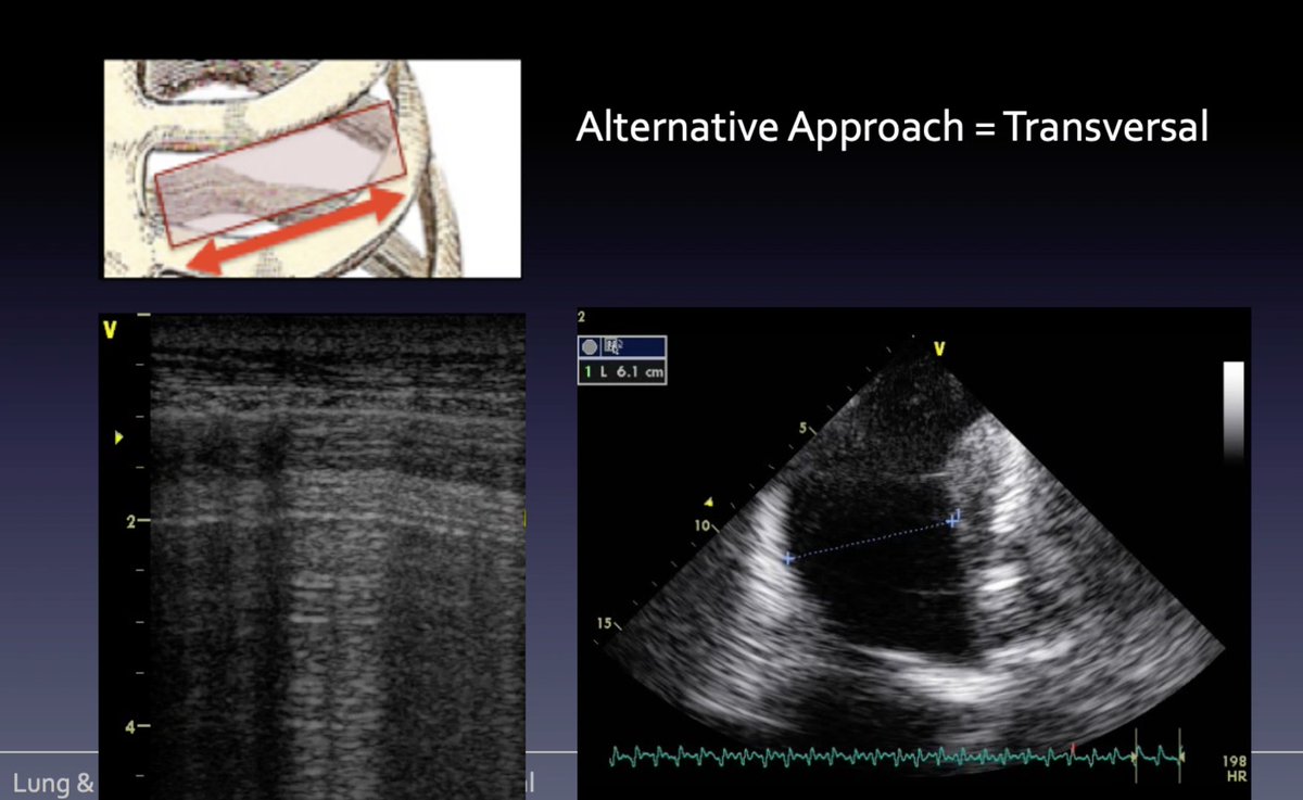
➡️longitudinal (standard) = transversal to ICS; smaller ICS = smaller window
➡️transversal (alternative) = aligned to ICS
Check
📰 bit.ly
📰 bit.ly
🧵Mongodi S tutorial #eSMART2021 #POCUS

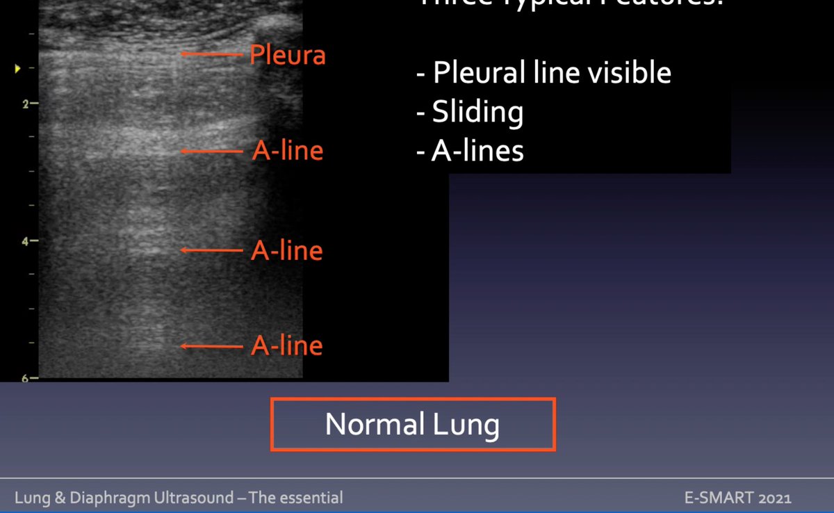
Tissue-Air interface in lung #ultrasound = artifacts normal or pathologic vs real images = no air = always pathologic. Normal LUS? 3 features
➡️visible pleural line
➡️pleural sliding (with ventilation)/sandy pattern in Mmode
➡️A-lines beneath pleura
🧵Mongodi S #eSMART2021 #POCUS
➡️visible pleural line
➡️pleural sliding (with ventilation)/sandy pattern in Mmode
➡️A-lines beneath pleura
🧵Mongodi S #eSMART2021 #POCUS
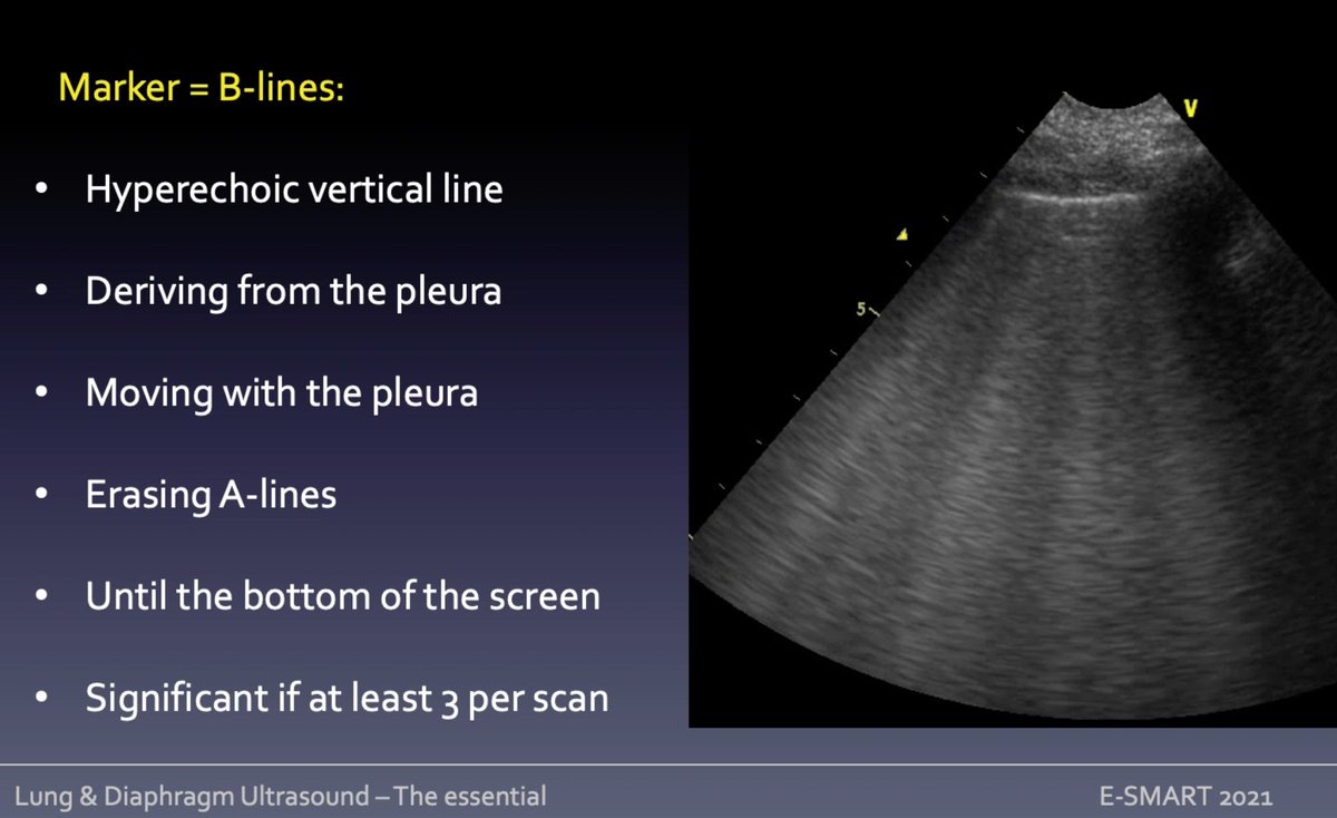
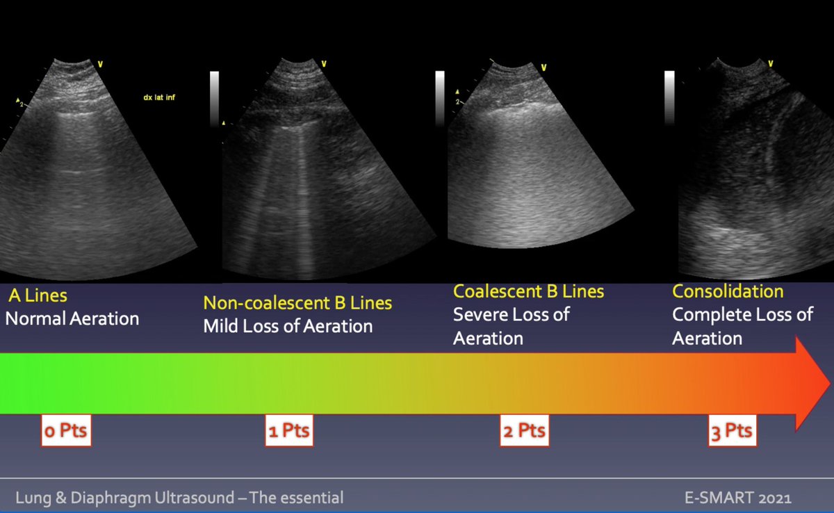
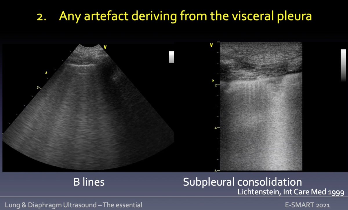
Alveolo-Interstitial syndrome at lung #POCUS defined by presence of 🅱️ lines hyperechoic vertical artifacts (arising from pleura/moving synchronous w it) = ⬆️lung density; at least 3 to be significant
⬆️n°/confluence =⬆️de-aeration
bit.ly
🧵Mongodi S #eSMART2021
⬆️n°/confluence =⬆️de-aeration
bit.ly
🧵Mongodi S #eSMART2021
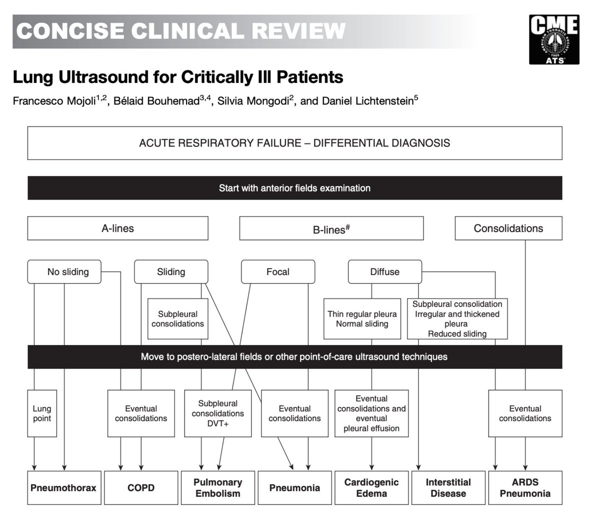
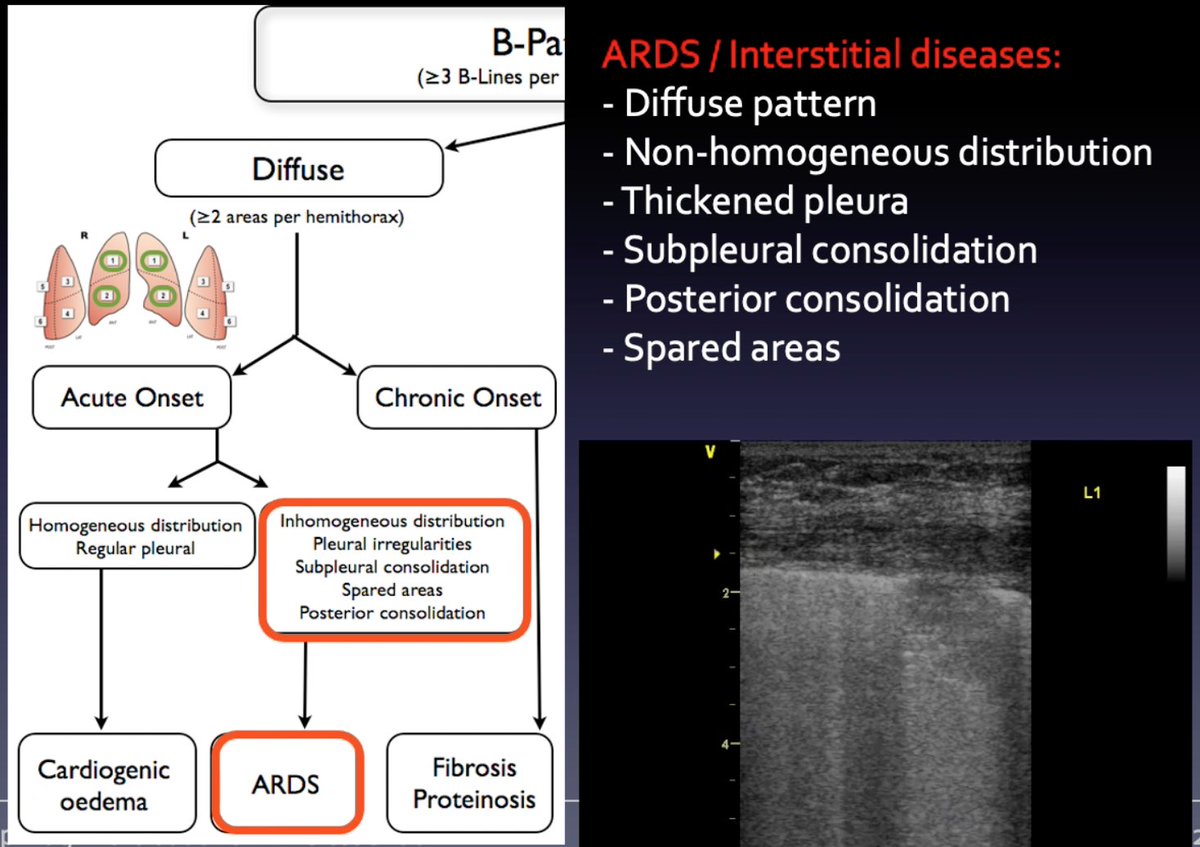
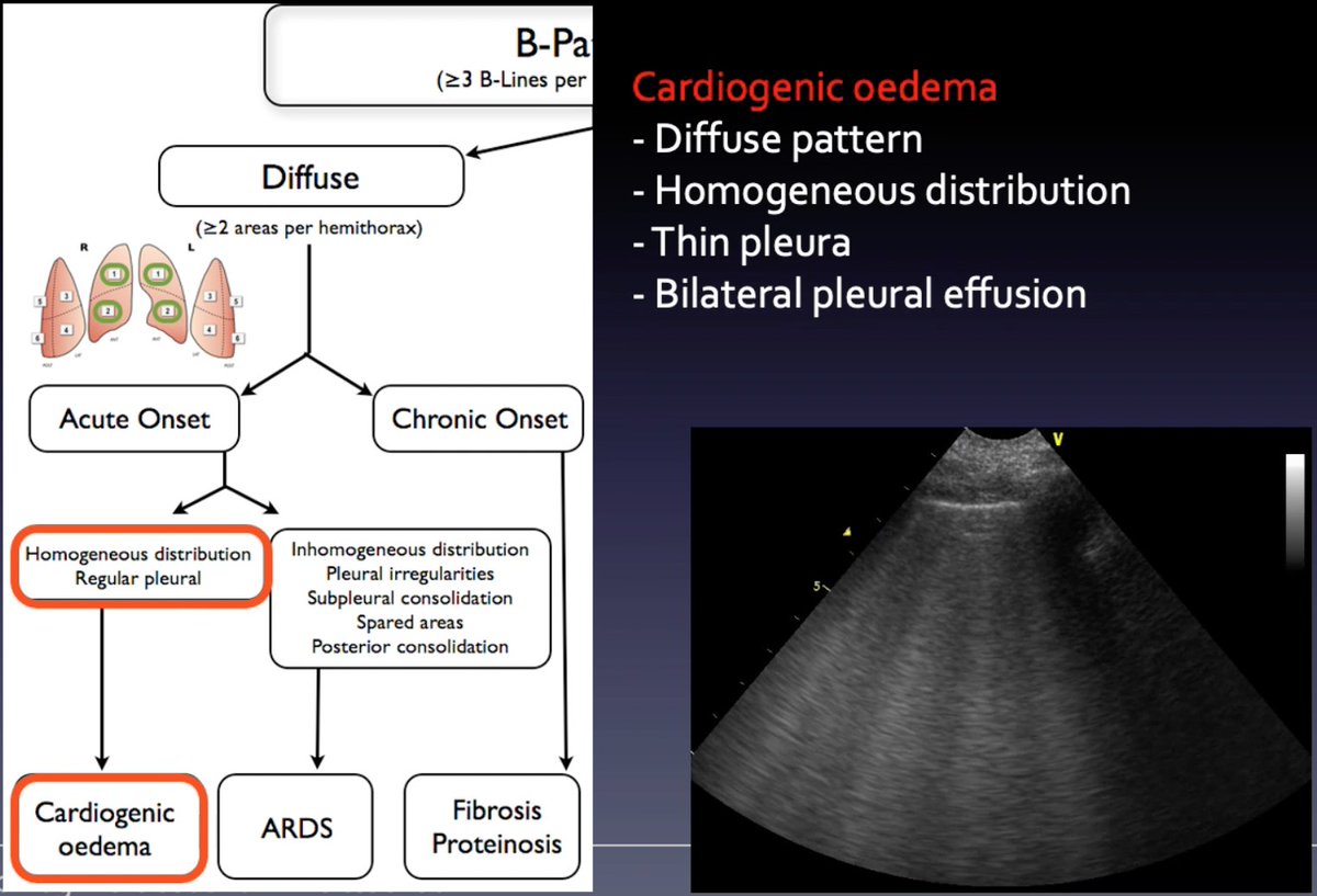
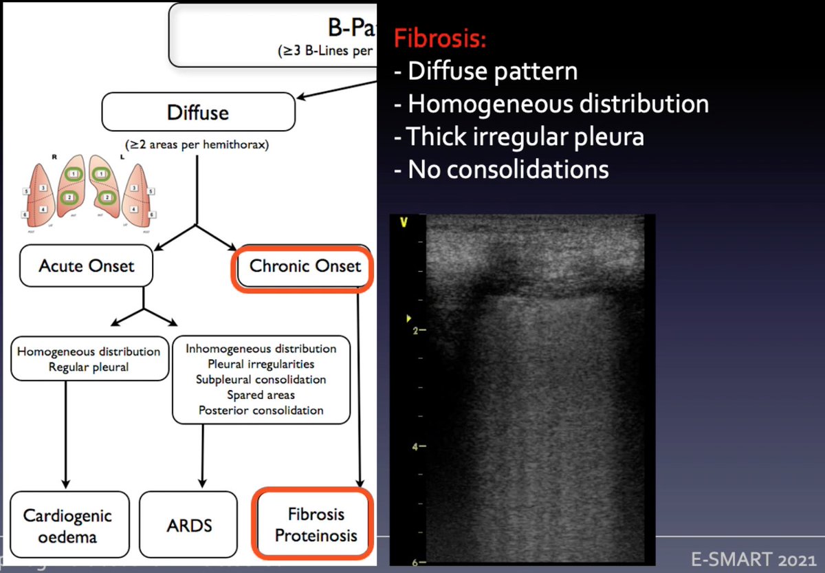
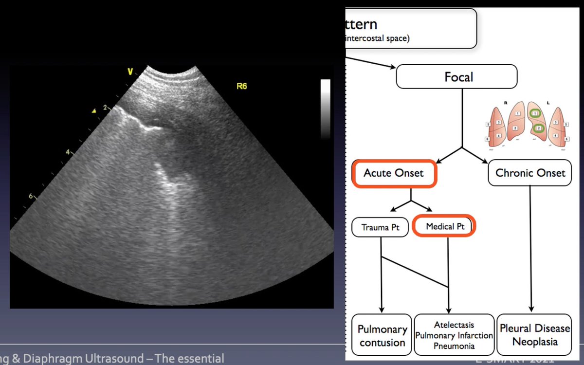
Integrate B lines w distribution/clinical signs/other #LUS signs
🅱️ pattern ≥3 B lines per ICS
➡️diffuse: ≥ 2 areas per hemithorax; acute onset? check distribution & pleura
➡️focal with acute onset? consider etiology trauma vs medical pt
🧵Mongodi S tutorial #eSMART2021 #POCUS
🅱️ pattern ≥3 B lines per ICS
➡️diffuse: ≥ 2 areas per hemithorax; acute onset? check distribution & pleura
➡️focal with acute onset? consider etiology trauma vs medical pt
🧵Mongodi S tutorial #eSMART2021 #POCUS
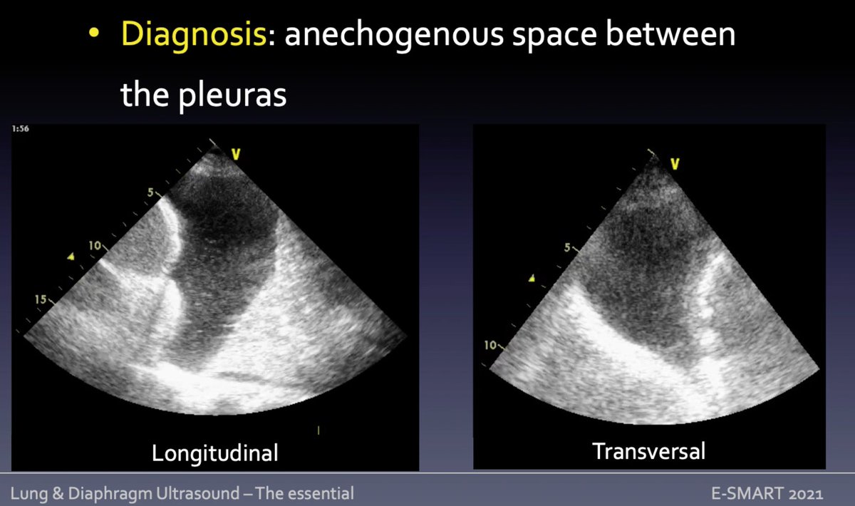
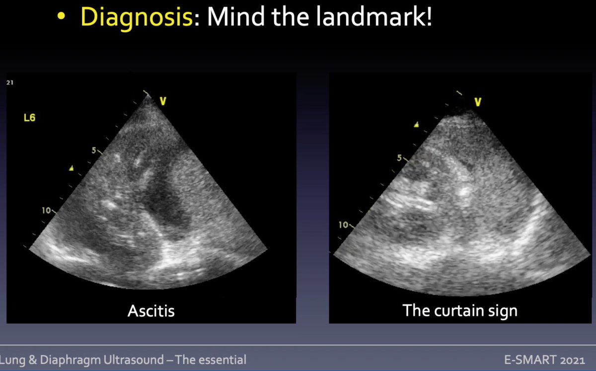
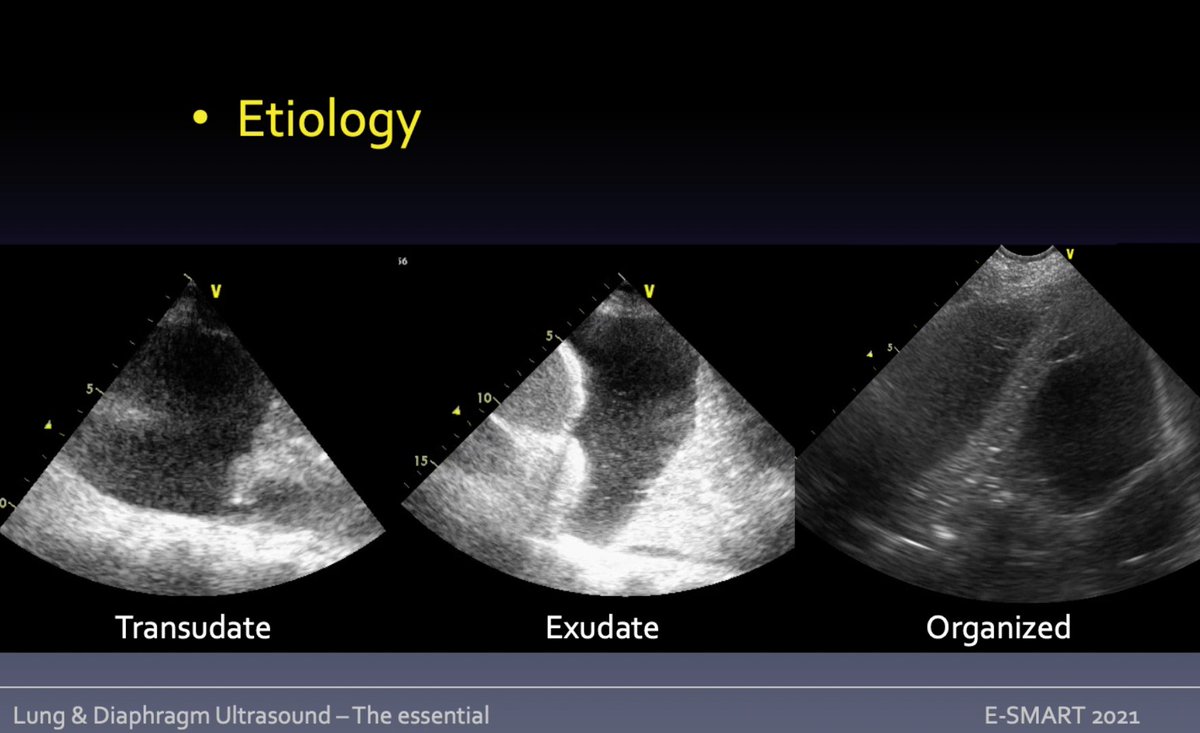
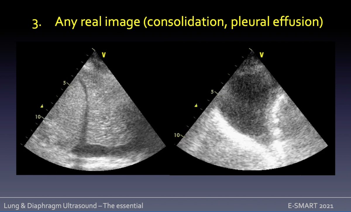
Pleural effusion = anechogenous space between pleuras. Is inside thorax? pleural vs abdominal fluid = aspect; curtain sign: aerated costophrenic recesses/peripheral bases.
➡️transudate: anechogenous/homogeneous
➡️SEC: inflammatory
➡️organized: septa
🧵Mongodi S #eSMART2021 #POCUS
➡️transudate: anechogenous/homogeneous
➡️SEC: inflammatory
➡️organized: septa
🧵Mongodi S #eSMART2021 #POCUS
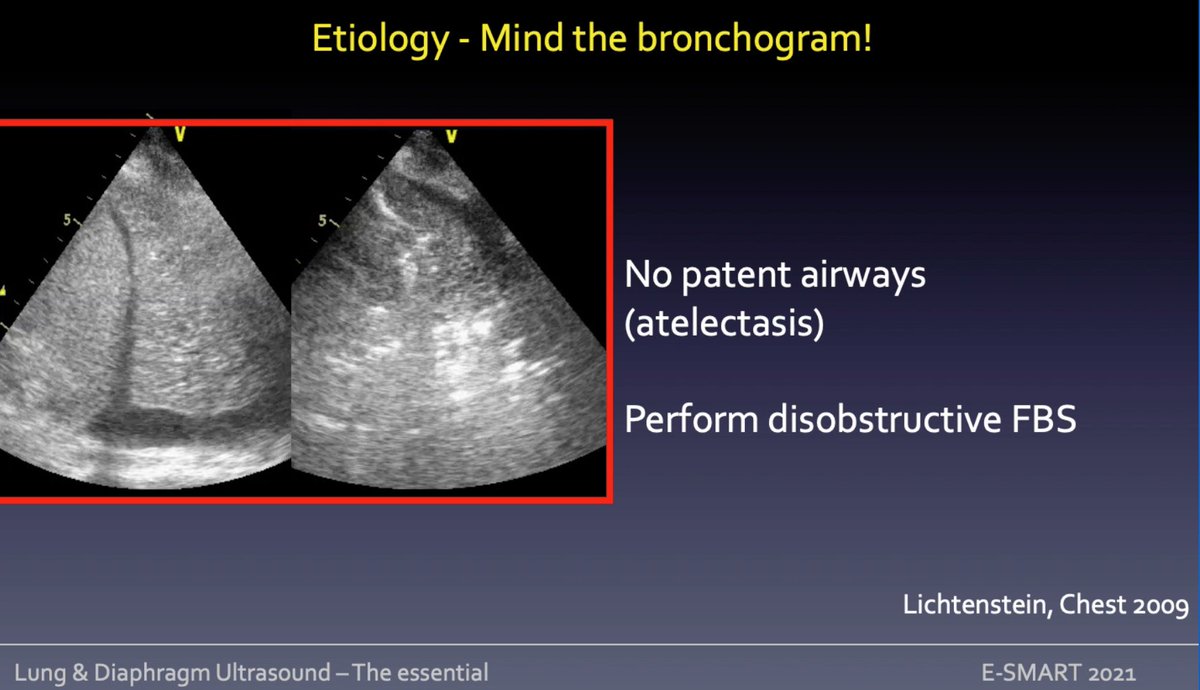
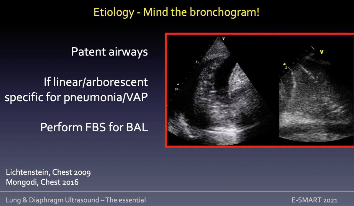
Consolidation at lung #ultrasound
1⃣tissue line pattern: lung appears as abdominal parenchyma = complete loss of aeration/lobe hepatization
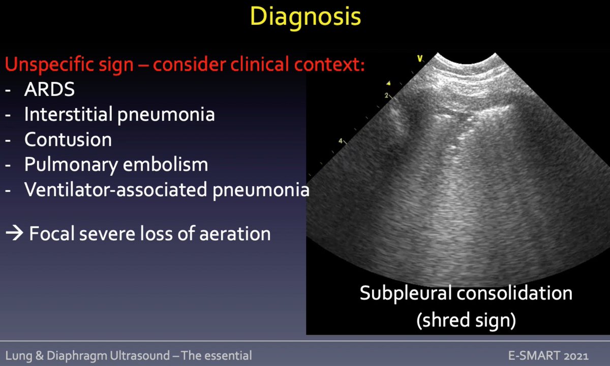
2⃣shred sign subpleural consolidation = echo poor region applied to pleura witch appear irregular
🧵 Mongodi S #eSMART2021 #POCUS #echofirst
1⃣tissue line pattern: lung appears as abdominal parenchyma = complete loss of aeration/lobe hepatization
2⃣shred sign subpleural consolidation = echo poor region applied to pleura witch appear irregular
🧵 Mongodi S #eSMART2021 #POCUS #echofirst

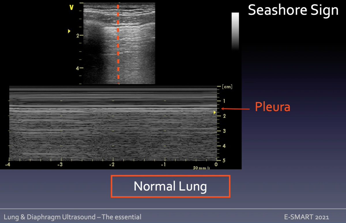
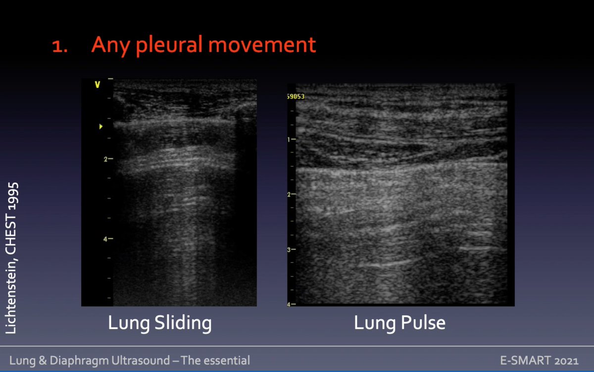
Pneumothorax at lung #ultrasound? easy to rule out if any
➡️pleural movement: sliding/pulse = NO PTX (you can confirm with M mode)
➡️artifact arising from visceral pleura ie B lines/consolidation
➡️real image ie effusion/consolidation
🧵Mongodi S #eSMART2021 #POCUS #echofirst
➡️pleural movement: sliding/pulse = NO PTX (you can confirm with M mode)
➡️artifact arising from visceral pleura ie B lines/consolidation
➡️real image ie effusion/consolidation
🧵Mongodi S #eSMART2021 #POCUS #echofirst
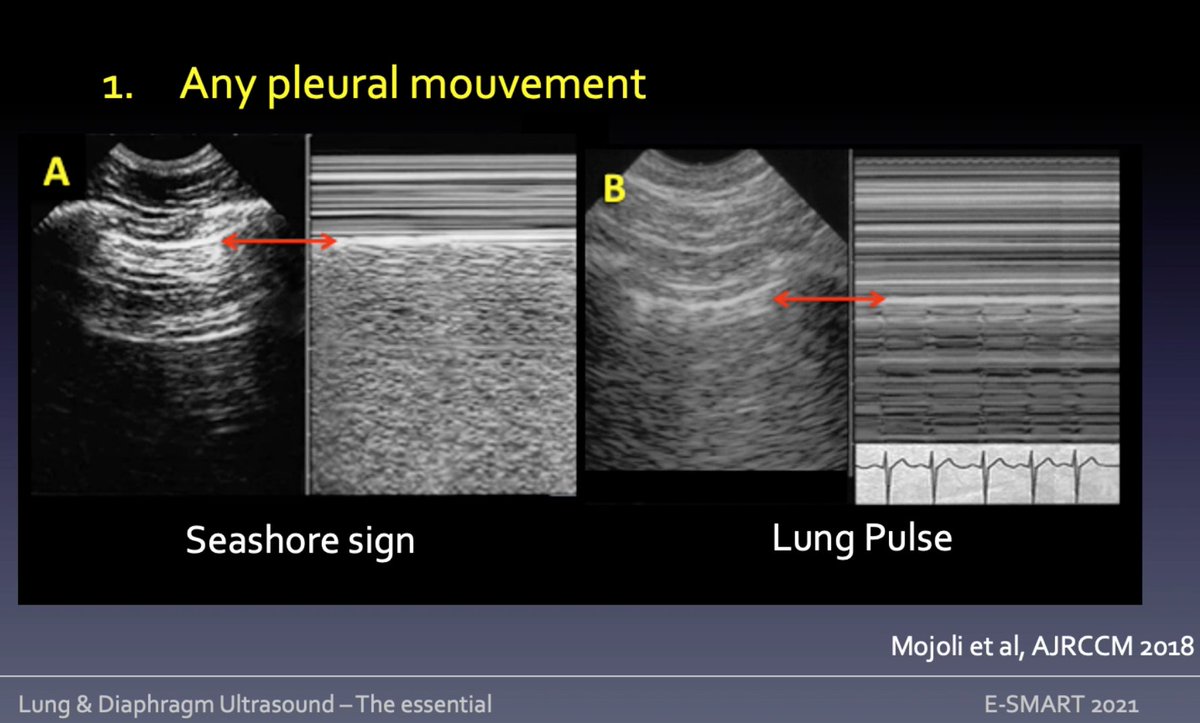
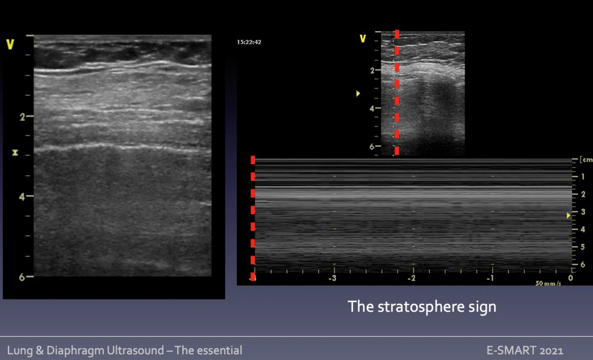
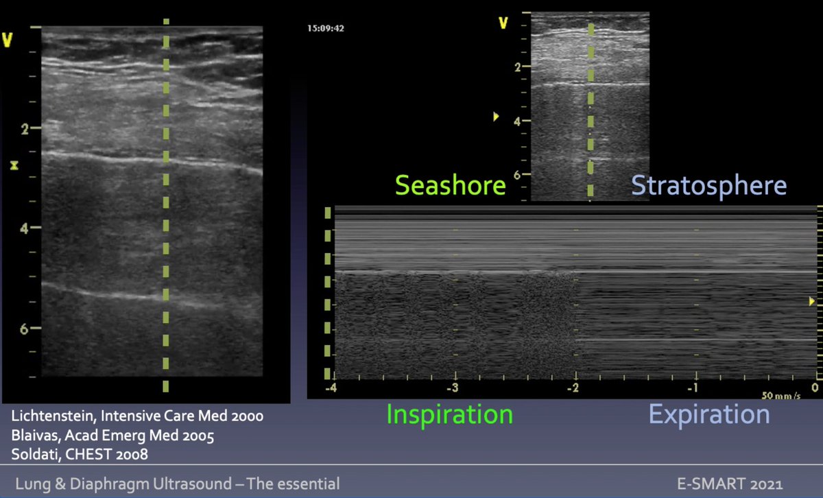
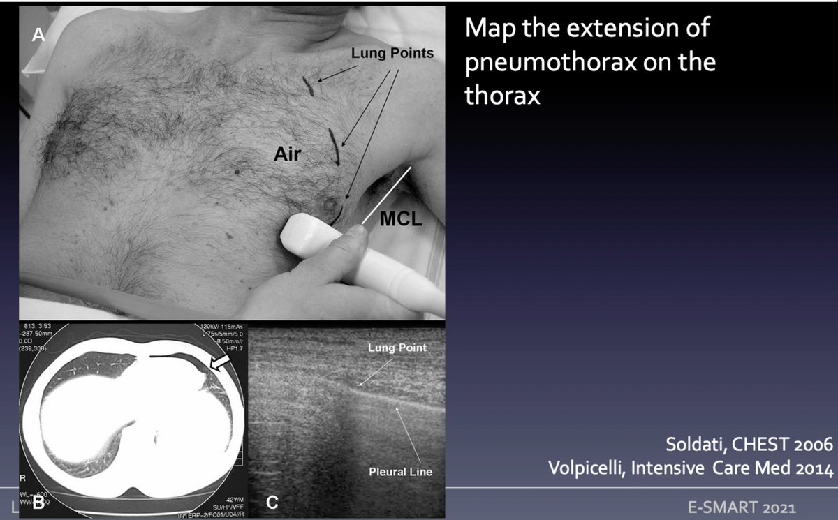
No pleural movement, static A pattern (confirmed by M mode: stratosphere sign): PTX? look for point where collapsed lung comes in touch w parietal pleura = lung point: switch seashore/stratosphere in M mode. No lung point? may be complete collapse!
🧵Mongodi S #eSMART2021 #POCUS
🧵Mongodi S #eSMART2021 #POCUS
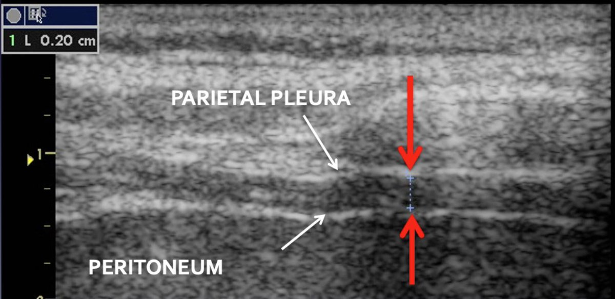
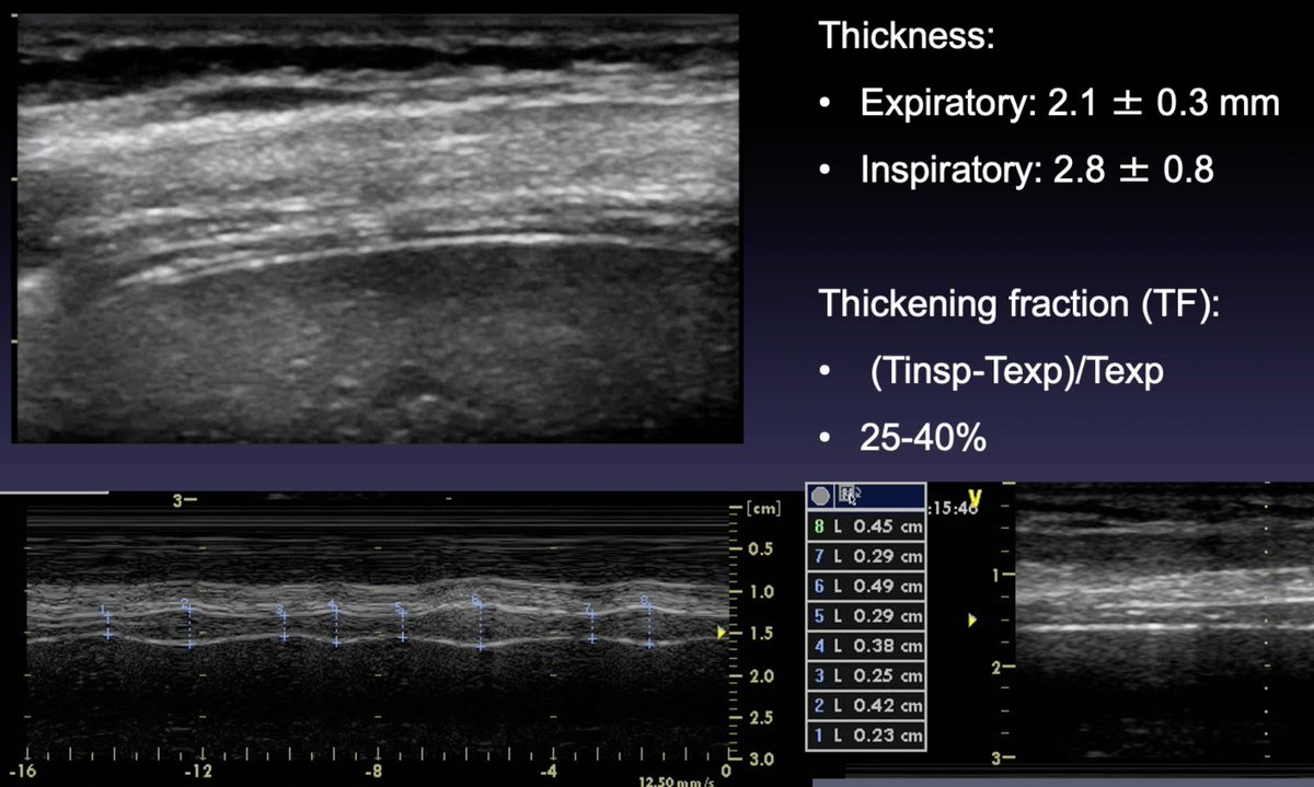
Diaphragm #POCUS assess
➡️excursion = displacement during inspiratory effort (affected by P on MV) + inspiratory/expiratory times (use anatomical M-Mode to align with direction of diaphragm excursion)
➡️thickness = variation during respiratory cycle + TF
🧵 Mongodi S #eSMART2021
➡️excursion = displacement during inspiratory effort (affected by P on MV) + inspiratory/expiratory times (use anatomical M-Mode to align with direction of diaphragm excursion)
➡️thickness = variation during respiratory cycle + TF
🧵 Mongodi S #eSMART2021


Loading suggestions...