

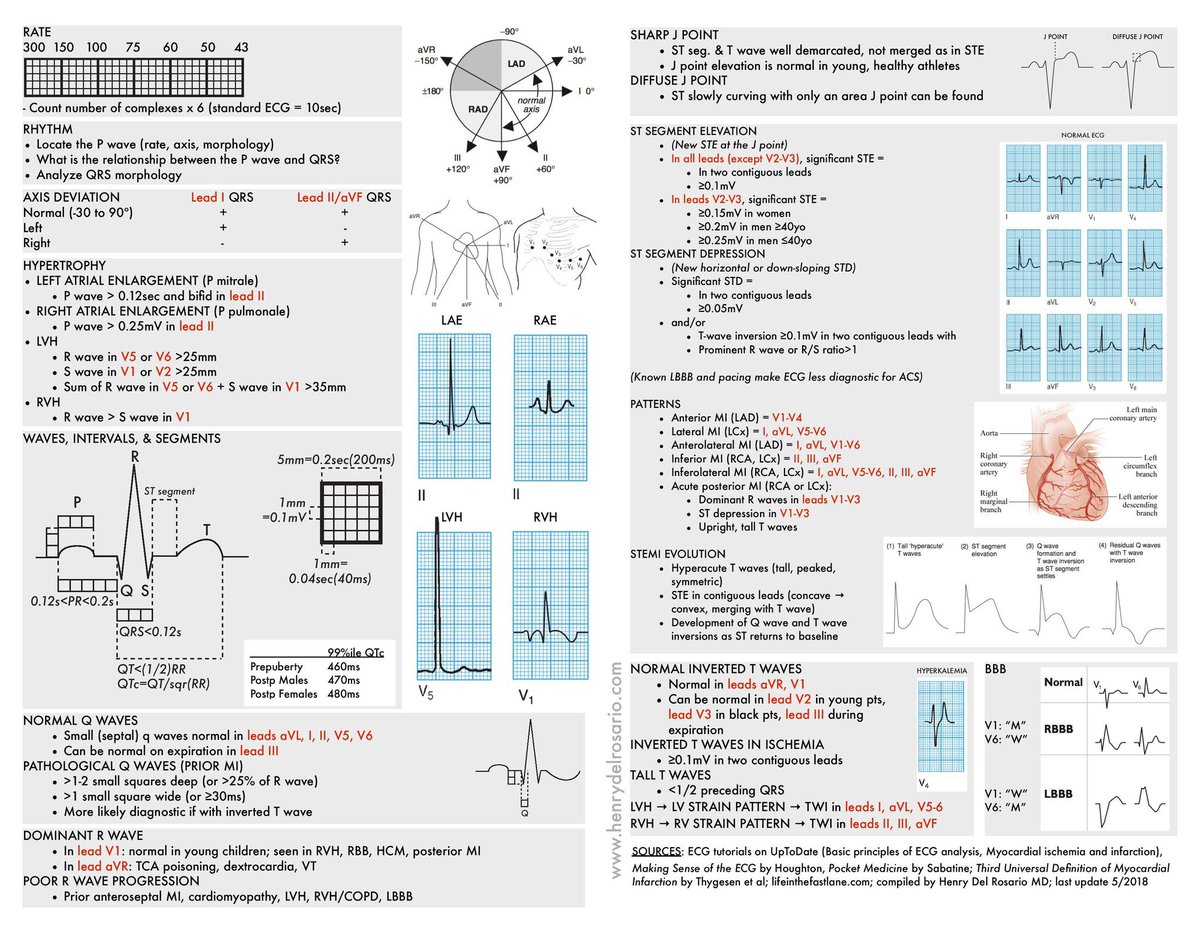
Let’s thread-together-ECG cheat sheets. Beginning from this masterpiece from @grepmeded
#EPeeps @SchakrabartiEP @ecgrhythms @abhishek_mbbs @syamkumarmd @mahishasur @DCR_Dr @anunay_cardio
@Hapa_EP @PaymardM @javadm20 @jeffrey_vinocur
@narrowQRS @seshadribalaji6 @rdschaller
#EPeeps @SchakrabartiEP @ecgrhythms @abhishek_mbbs @syamkumarmd @mahishasur @DCR_Dr @anunay_cardio
@Hapa_EP @PaymardM @javadm20 @jeffrey_vinocur
@narrowQRS @seshadribalaji6 @rdschaller
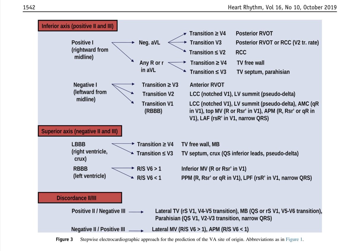
Idiopathic VT : 12-lead ECG to predict SOO
heartrhythmjournal.com
Andres Enriquez, Adrian Baranchuk, David Briceno, Luis Saenz, Fermin Garcia
heartrhythmjournal.com
Andres Enriquez, Adrian Baranchuk, David Briceno, Luis Saenz, Fermin Garcia

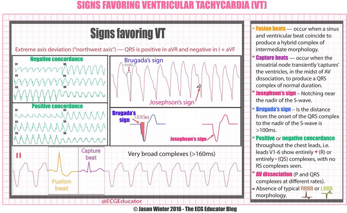
ECG signs favouring VT
(Courtesy Jason Winter,2016)
(Courtesy Jason Winter,2016)
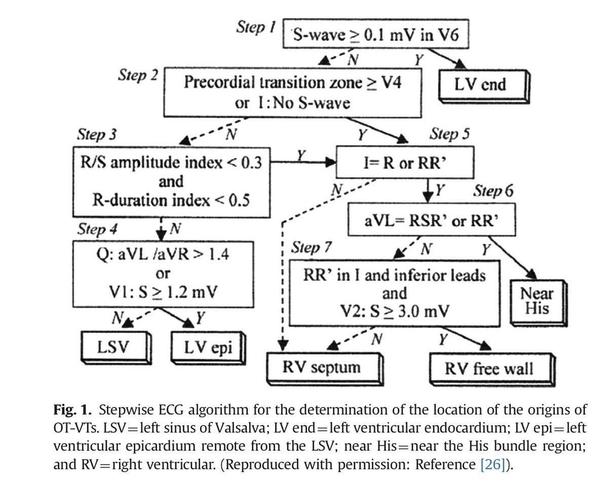
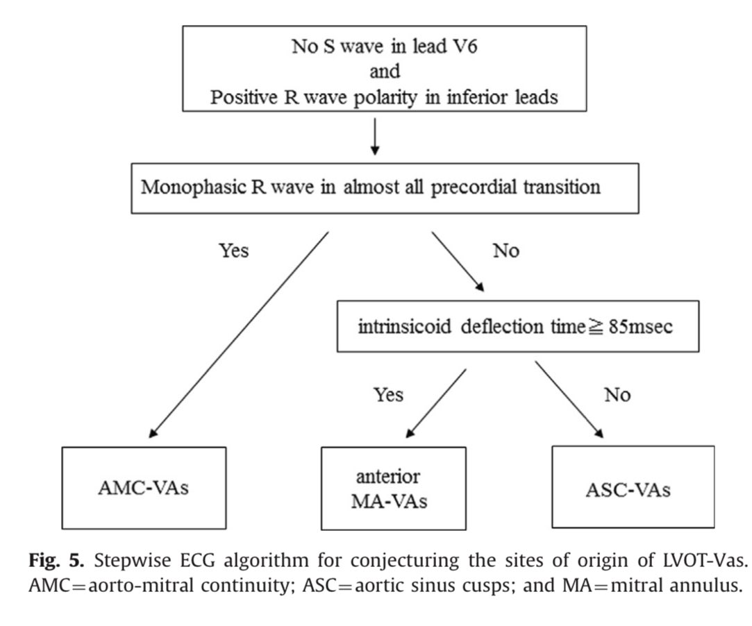
IDIOPATHIC OT- VT: SOO
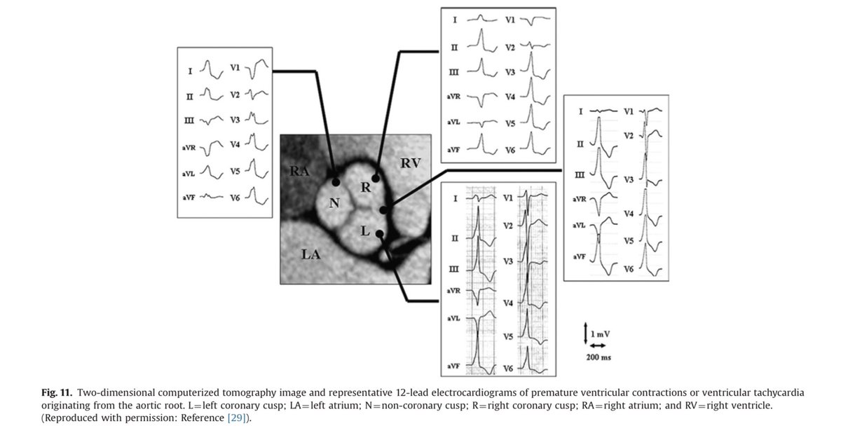
R or L?
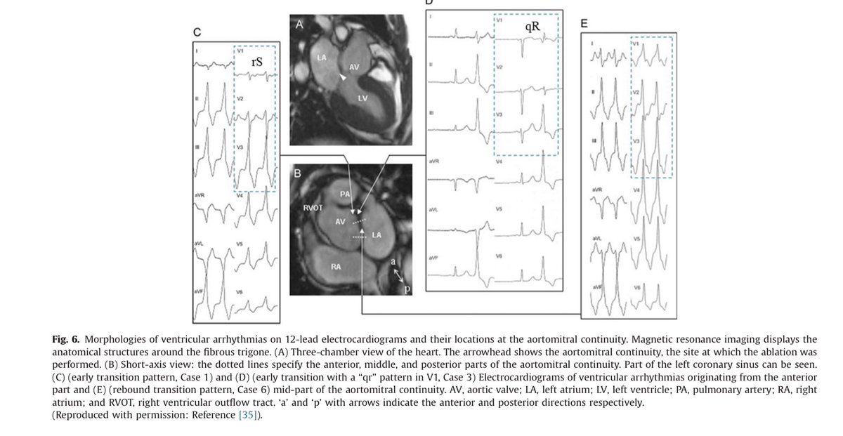
AMC or ASC or MA?
K. Kumagai / Journal of Arrhythmia 30
(2014) 211-221
R or L?
AMC or ASC or MA?
K. Kumagai / Journal of Arrhythmia 30
(2014) 211-221
Mechanism of aberrant conduction
Action potential, beautiful!
D/D Eisenmenger Syndrome (DrRajesh)
Fascicular VT or RBBB @rajivasr
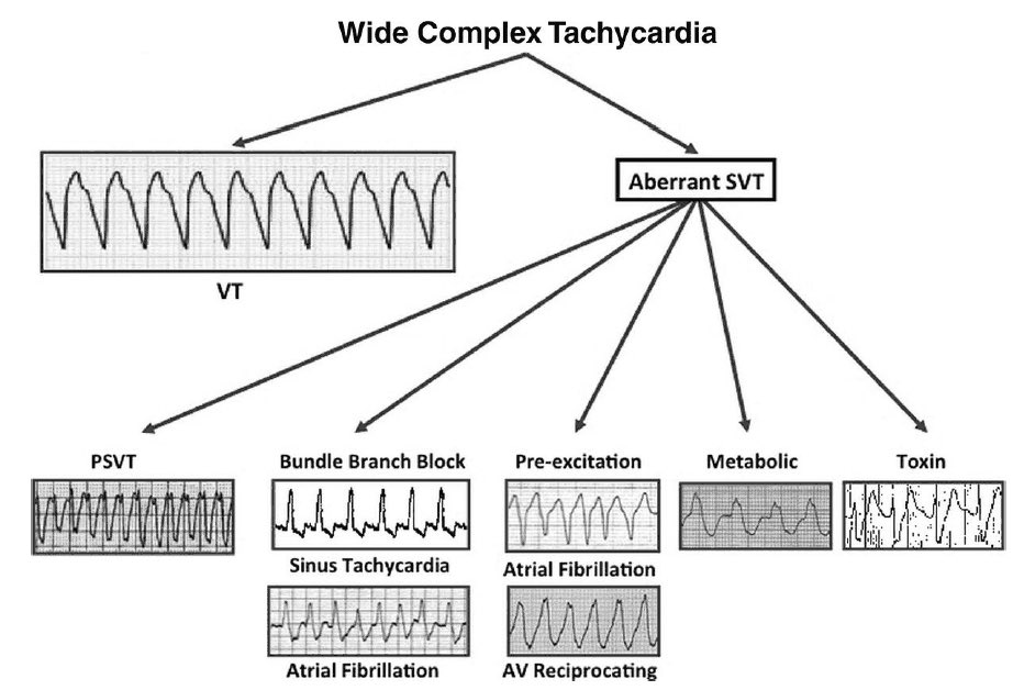
WCT D/D
@ManualOMedicine
@ManualOMedicine
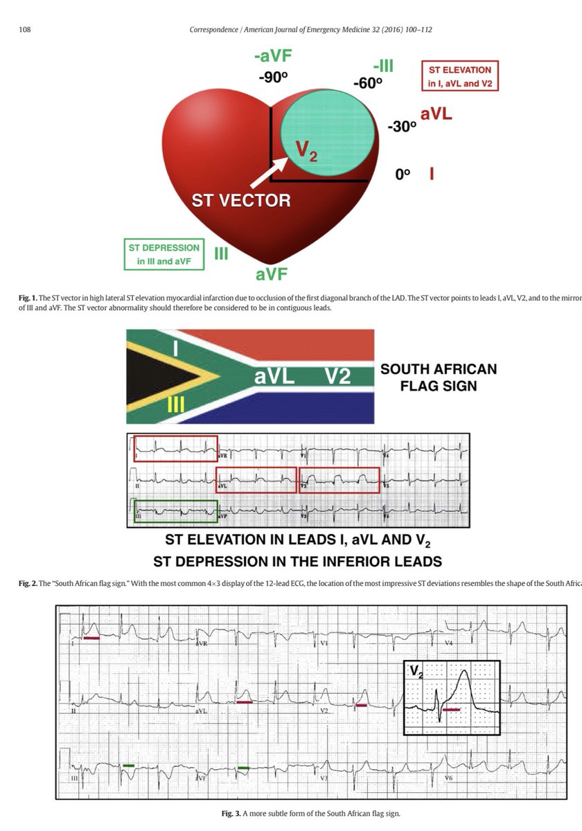
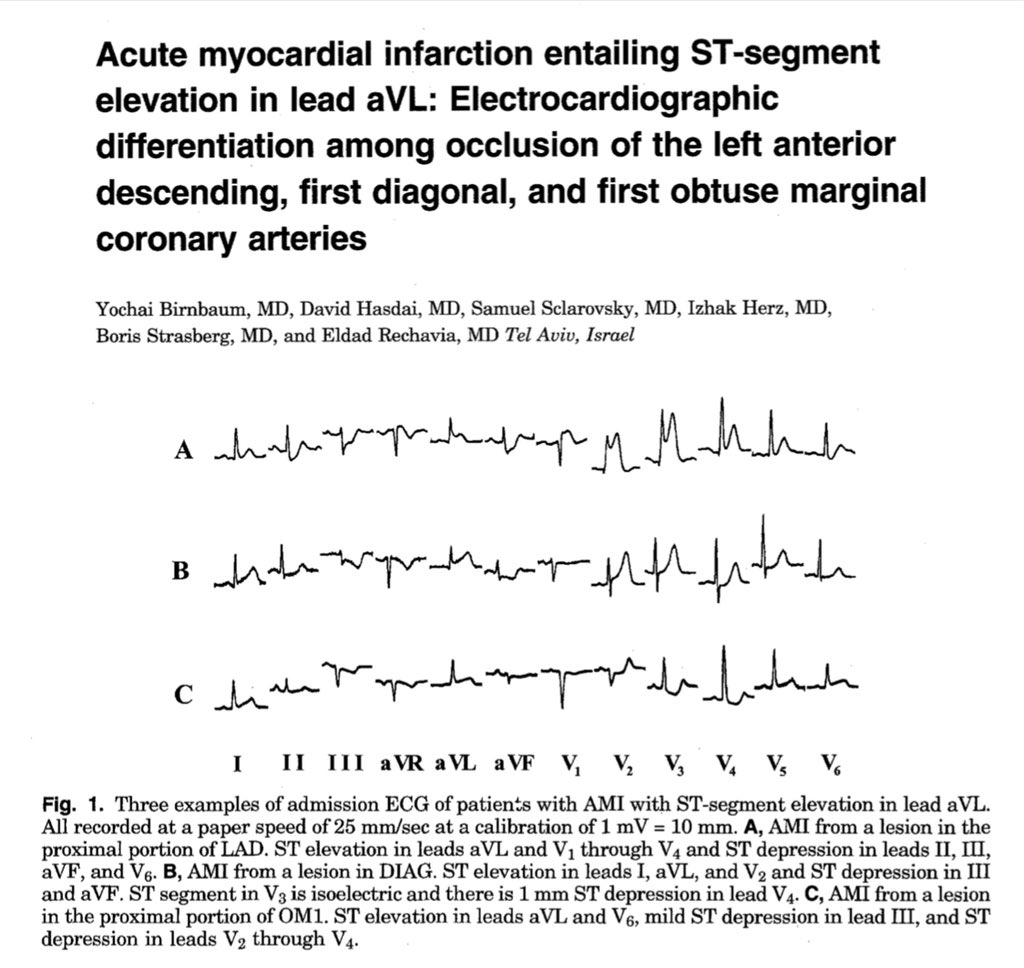
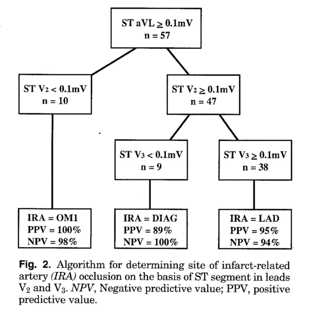
South African Flag Sign:
ECG features of Acute High Lateral wall MI due to D1 occlusion.
ECG features of Acute High Lateral wall MI due to D1 occlusion.


ECG and other markers of risk for Sudden Cardiac Arrest @EPeeps_Bot
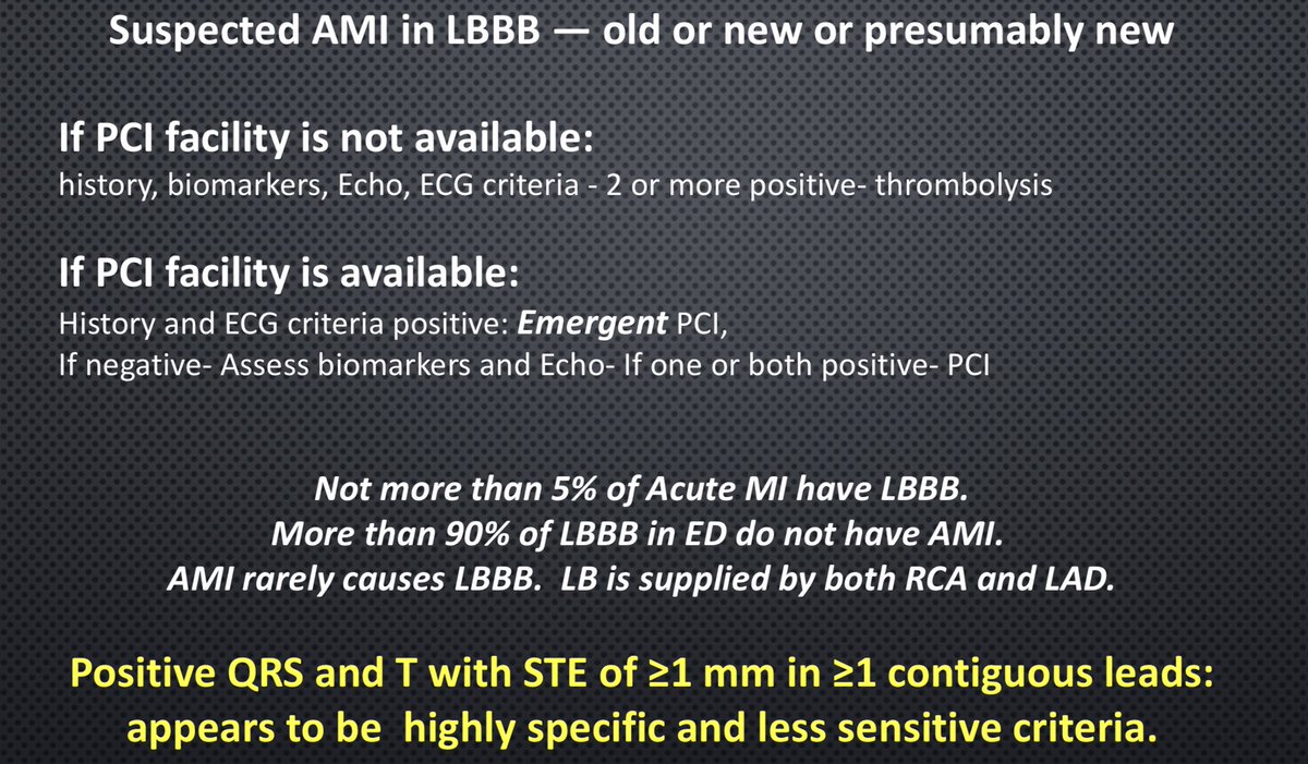
LBBB: Is it AMI?
Depends on how do you like to treat! By Thrombolysis or PCI?
A useful (?) algorithm below
@EPeeps_Bot
Depends on how do you like to treat! By Thrombolysis or PCI?
A useful (?) algorithm below
@EPeeps_Bot
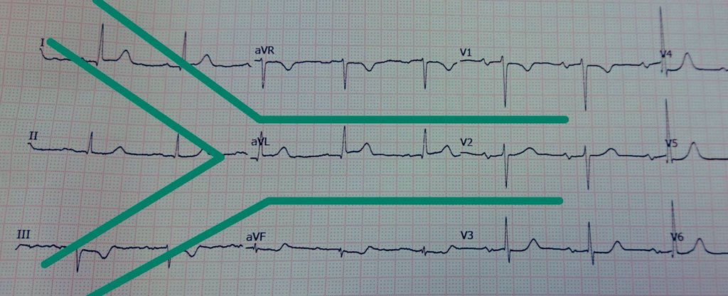
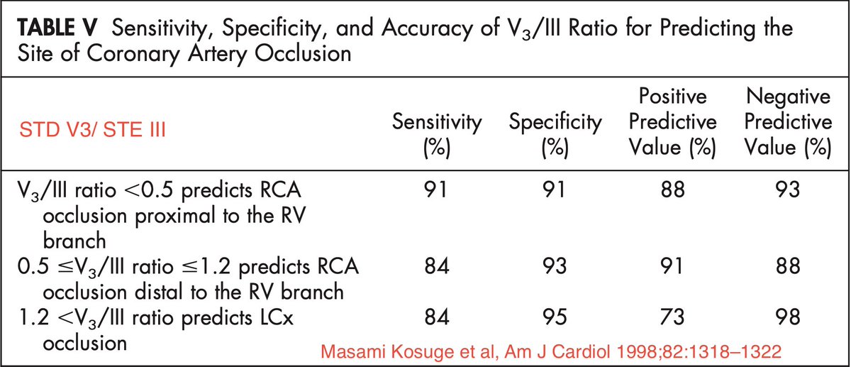
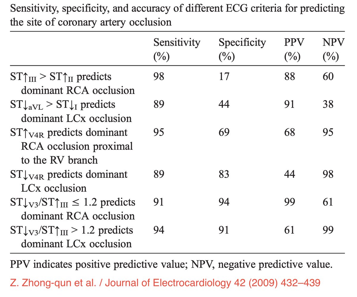
IWMI: IRA - RCA or LCX
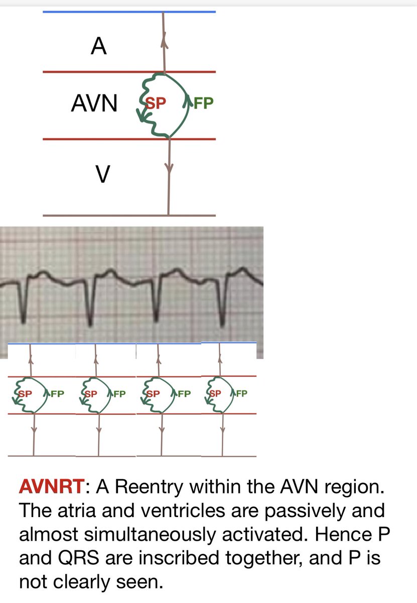
AVNRT:
Few questions- all mean the same!
Why P is not clearly seen?
Or
Why P is buried within QRS?
Or
Why is it a ‘Short RP’ tachycardia?
Single answer:
Atria and ventricles activated near simultaneously!
Here it’s how:
#ECG
Few questions- all mean the same!
Why P is not clearly seen?
Or
Why P is buried within QRS?
Or
Why is it a ‘Short RP’ tachycardia?
Single answer:
Atria and ventricles activated near simultaneously!
Here it’s how:
#ECG
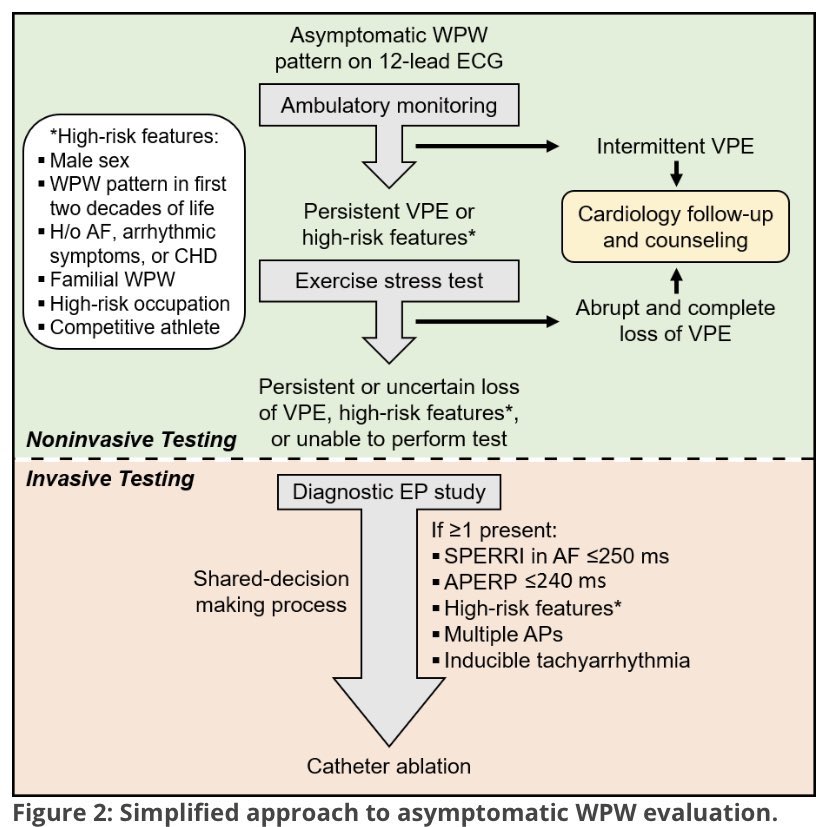
Asymptomatic WPW pattern: Risk stratification
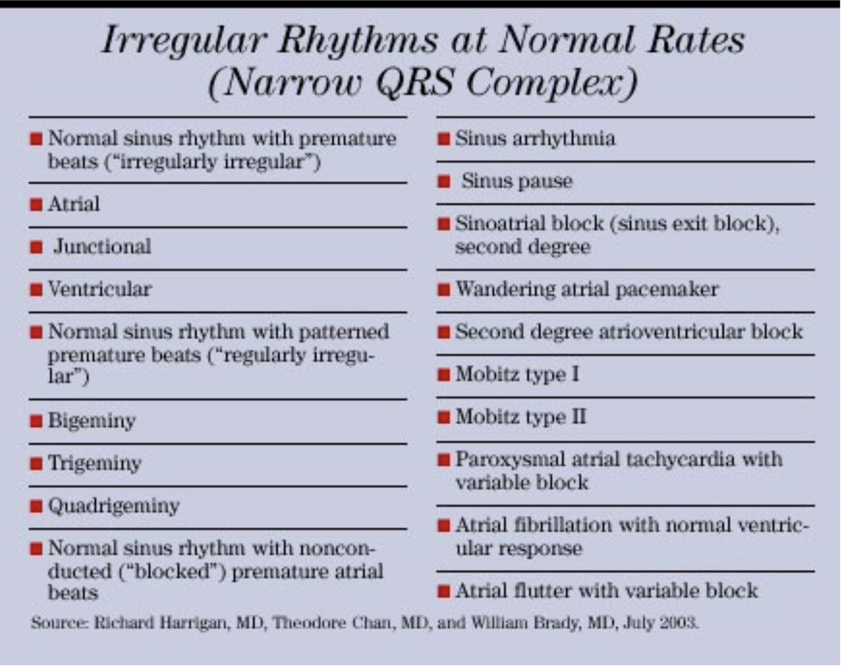
Irregular Rhythms at Normal Rates (Narrow QRS Complex)
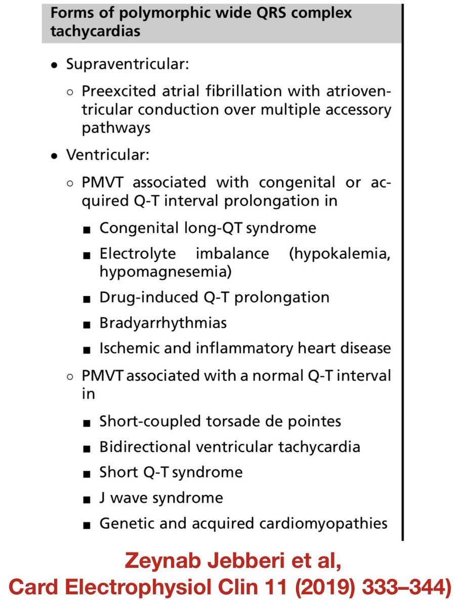
Polymorphic Wide QRS Complex Tachycardia Differential Diagnosis
(Commonest cause: Ventricular origin) #ECG @EPeeps_Bot
(Commonest cause: Ventricular origin) #ECG @EPeeps_Bot
ECG Books #ECG
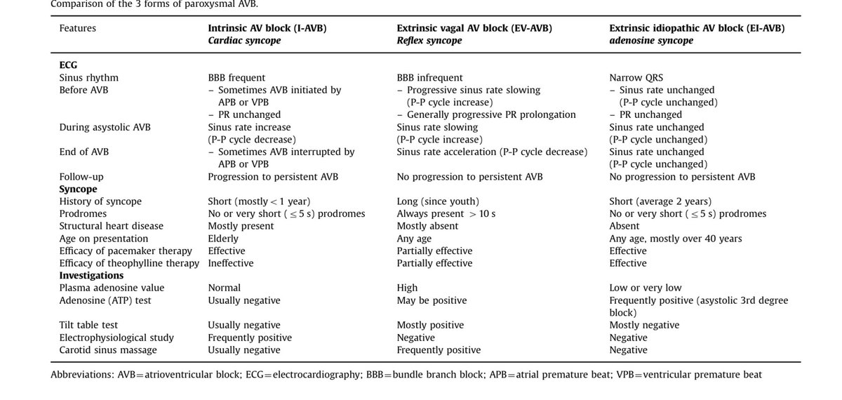
Syncope and paroxysmal atrioventricular block: Milena Aste, Michele Brignole, Journal of Arrhythmia 33 (2017) 562–567 #ECG @EPeeps_Bot
20Y healthy medico, health screening
Morphology of PVC originating from near the His Bundle
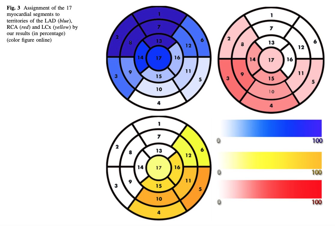
IRA and CORONARY ARTERY TERRITORY:
Often some anatomical variability.
Occasionally previous occlusions with collaterals.
Hence the concept of IRA is a conjecture.
AHA statement and Real world data.
(P Donato et al) @EPeeps_Bot #ECG
Often some anatomical variability.
Occasionally previous occlusions with collaterals.
Hence the concept of IRA is a conjecture.
AHA statement and Real world data.
(P Donato et al) @EPeeps_Bot #ECG


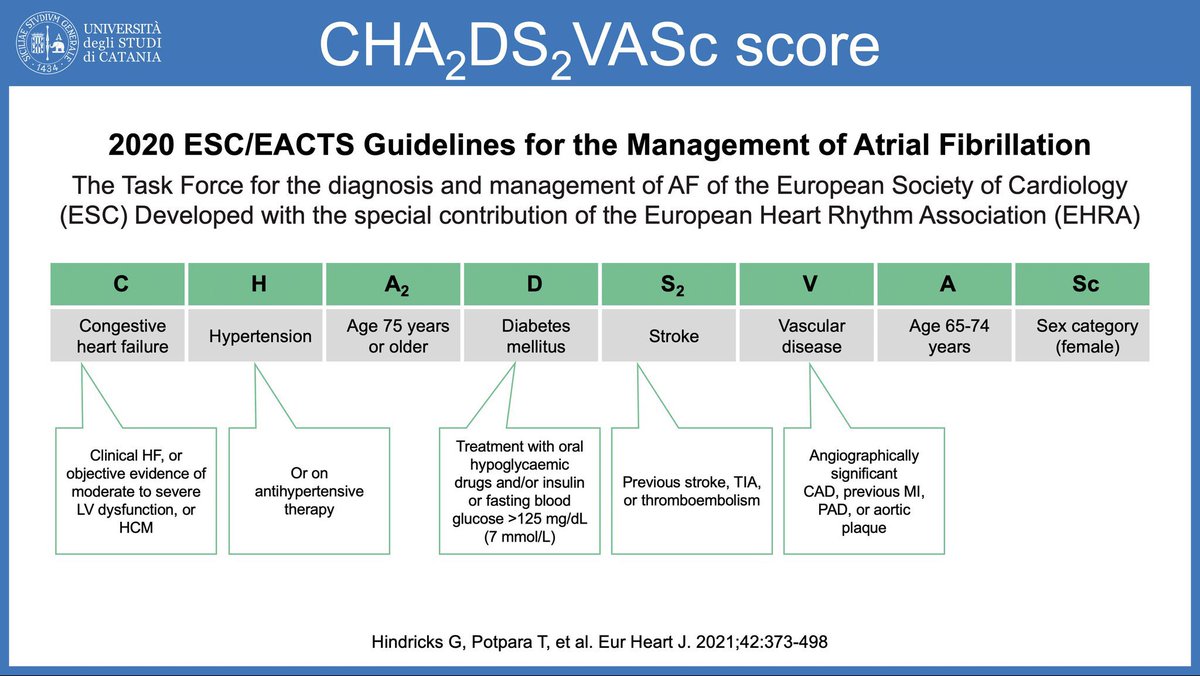
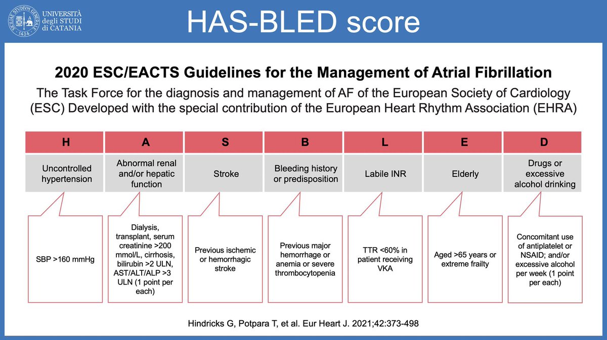
SPAF:
Stroke and Bleeding Scores
Stroke and Bleeding Scores
Loading suggestions...