

I’m too bright for this scan..so bright, it hurts! A tweetorial.
60 year old with cirrhosis & klebsiella bacteremia 👇 status epilepticus & hepatic encephalopathy
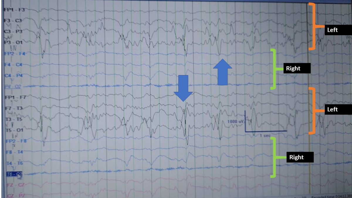
EEG- left hemispheric status epilepticus followed by PLEDS. CT is reported as subdural hygroma.
60 year old with cirrhosis & klebsiella bacteremia 👇 status epilepticus & hepatic encephalopathy
EEG- left hemispheric status epilepticus followed by PLEDS. CT is reported as subdural hygroma.
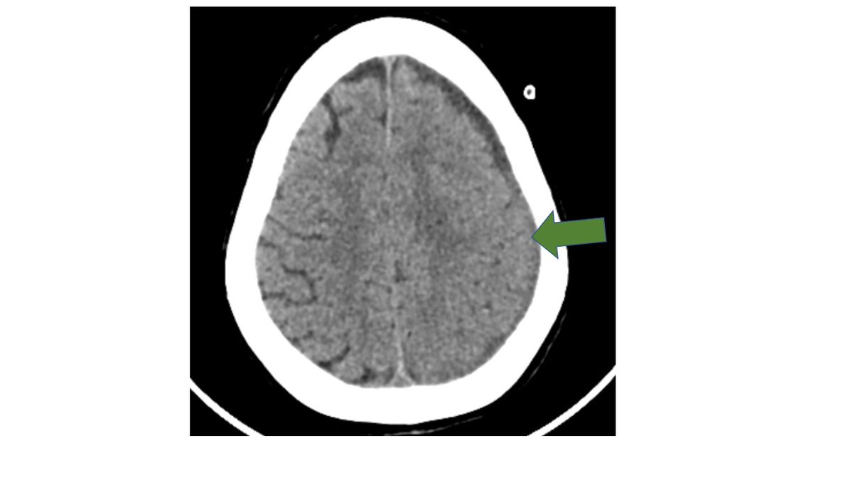
EEG shows left hemispheric rhythmic discharges s/o focal status epilepticus. A small subdural hygroma can't possibly cause this much trouble?
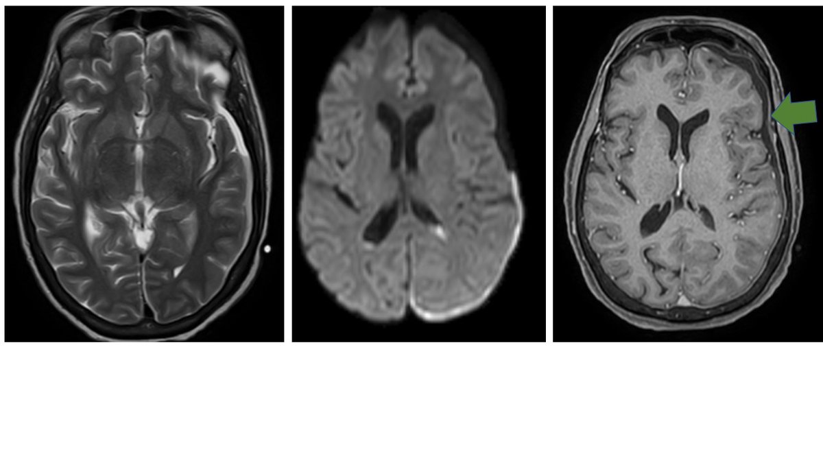
MRI shows layering in the subdural with a BRIGHT signal on Diffusion imaging as well as mild pachymeningeal enhancement- looks suspicious-Common infectious diseases of the central nervous system014clinical features and imaging characteristics ncbi.nlm.nih.gov
Clinical findings disproportionate to the imaging findings or underlying bacteremia, sepsis or immunosuppression should arouse the suspicion of a subdural empyema. Main d/ds are a subdural hygroma or hematoma. All of them can be hypodense to isodense to brain on NECT.
Non contrast MRI also can look identical
However contrast administration demonstrates diffuse meningeal enhancement in both CT and MR
Restricted diffusion of pus also differentiates subdural empyema from subdural hygroma, or hematoma.
However contrast administration demonstrates diffuse meningeal enhancement in both CT and MR
Restricted diffusion of pus also differentiates subdural empyema from subdural hygroma, or hematoma.
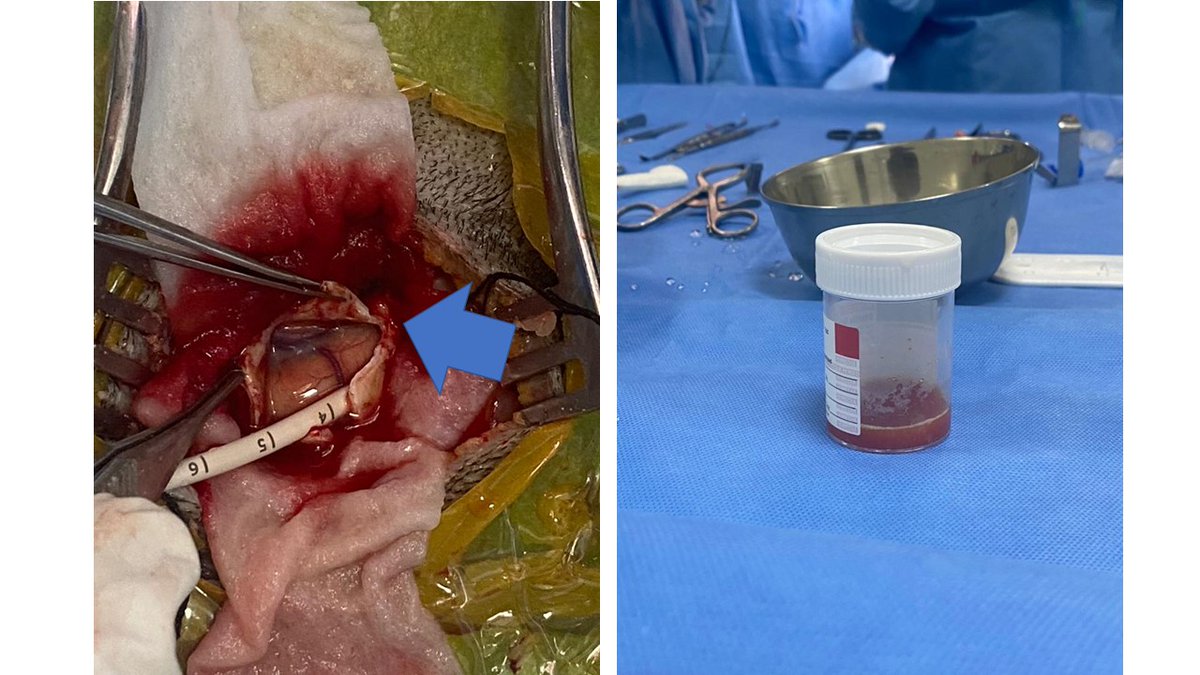
Fortunately, the neurosurgeon jumps into this nasty picture and drains out......pus. It's indeed a Subdural empyema........When in doubt, drain out !
Loading suggestions...