

It's important to be familiar with normal post-op imaging findings following cranial surgeries with different surgical techniques such as :
burr holes, craniotomy, craniectomy,and cranioplasty.
I show you different normal post-op images in a row as remarks. I hope you like it👍
burr holes, craniotomy, craniectomy,and cranioplasty.
I show you different normal post-op images in a row as remarks. I hope you like it👍

Remark 1
After craniotomy, contrast ++ seen both in CT&MRI, lasting longer at MR .
The dura mater enhances in a smooth linear pattern as soon as 9 hours after surgery,& enhancement can last as long as 40 years!
It almost always occurs in the portion of dura mater deep to flap
After craniotomy, contrast ++ seen both in CT&MRI, lasting longer at MR .
The dura mater enhances in a smooth linear pattern as soon as 9 hours after surgery,& enhancement can last as long as 40 years!
It almost always occurs in the portion of dura mater deep to flap

Remark 2
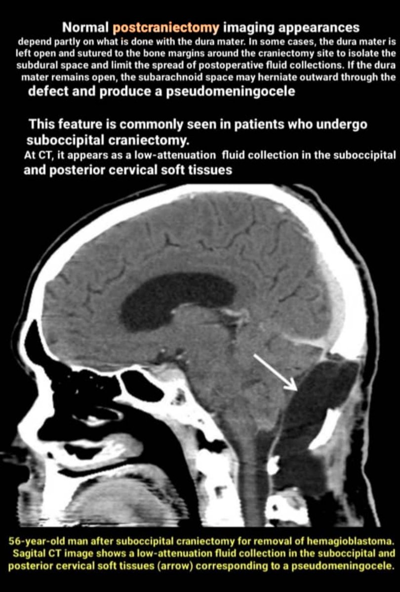
Craniectomy is the removal of a portion of the skull without subsequent replacement of the bone. It may be performed to remove an infected bone flap from previous craniotomy or a tumor that has infiltrated the calvaria. It may perform as part of a suboccipital approach
Craniectomy is the removal of a portion of the skull without subsequent replacement of the bone. It may be performed to remove an infected bone flap from previous craniotomy or a tumor that has infiltrated the calvaria. It may perform as part of a suboccipital approach
Remark 3
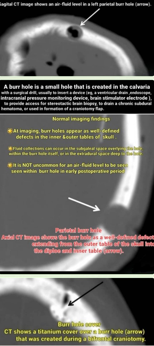
A burr hole is a small hole that is created in the calvaria with a surgical drill, usually to insert a device (eg, a ventricular drain or shunt catheter, ..), provide access for stereotactic brain biopsy, to drain a chronic SDH, or used in formation of a craniotomy flap
A burr hole is a small hole that is created in the calvaria with a surgical drill, usually to insert a device (eg, a ventricular drain or shunt catheter, ..), provide access for stereotactic brain biopsy, to drain a chronic SDH, or used in formation of a craniotomy flap
Remark 4
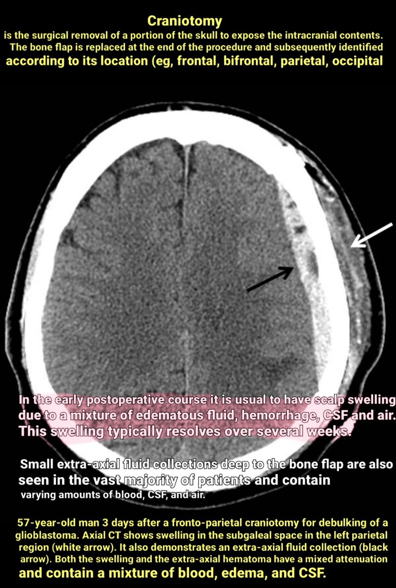
Craniotomy is the surgical removal of a portion of the skull to expose the intracranial contents. The bone flap is replaced at the end of the procedure and subsequently identified according to its location (eg, frontal, bifrontal, parietal, occipital).
Craniotomy is the surgical removal of a portion of the skull to expose the intracranial contents. The bone flap is replaced at the end of the procedure and subsequently identified according to its location (eg, frontal, bifrontal, parietal, occipital).
Remark 5
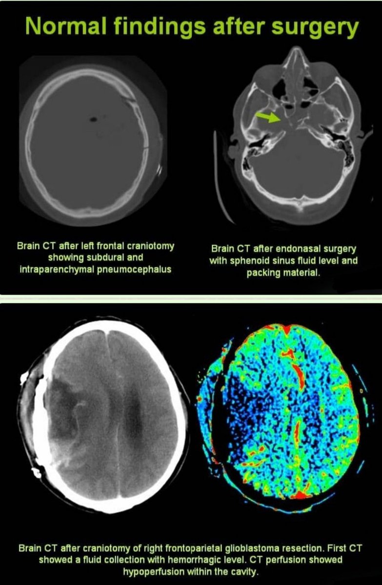
Two additional examples👇👇👇
Two additional examples👇👇👇
Loading suggestions...