

🧵1/16 Approach to reading a head CT: (1) Compare the anatomy of the images in front of you with a “normal” reference (in your head); (2) Look for specific abnormalities based on the clinical question/patient presentation/patient history. #radres #MedEd #futureradres #radiology

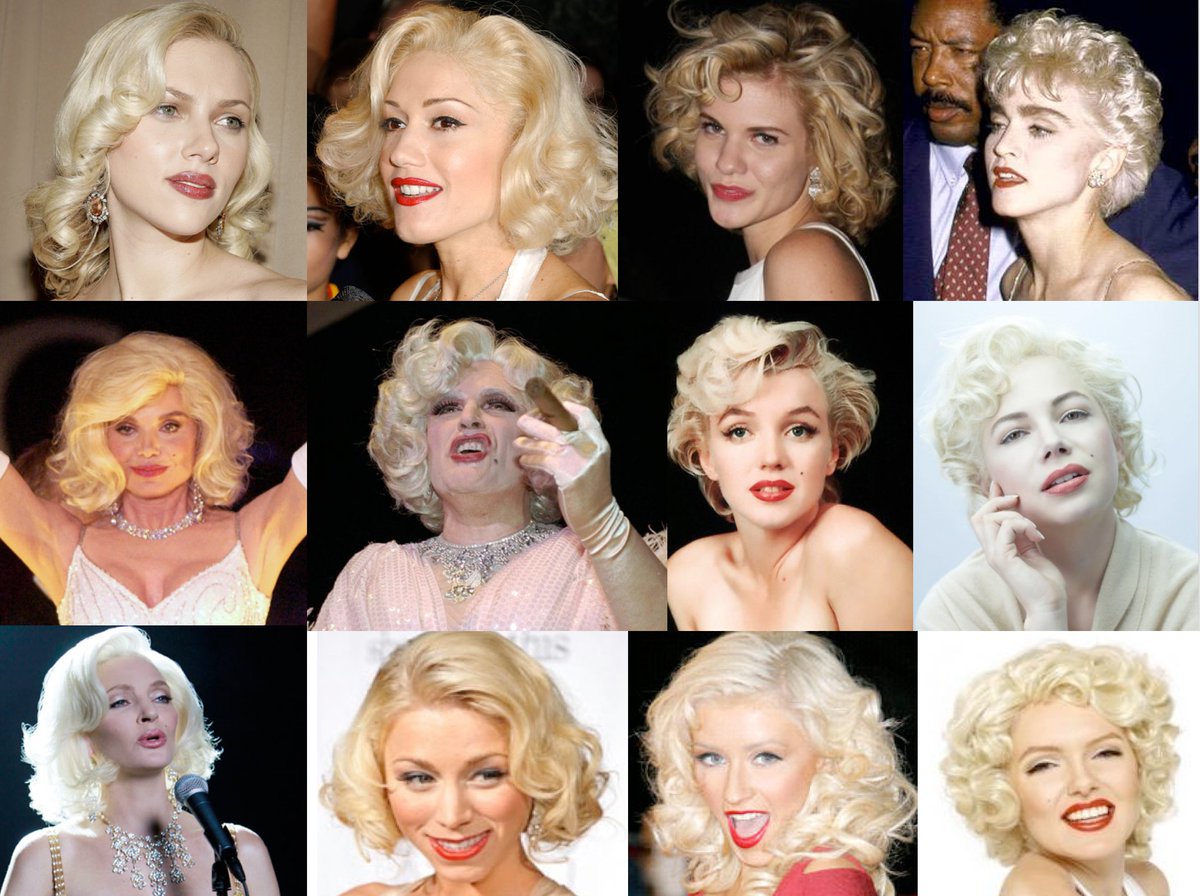
🧵2/16 Having a “normal” reference. You develop a “normal” database in your head by reviewing a lot of imaging studies, both normal and abnormal. With time, your brain will subconsciously raise a “red flag” when something looks “off” Like finding “real” Marilyn without effort!!
🧵3/16 Use window leveling and different imaging kernels to help with review of normal versus abnormal anatomy. #radres #MedEd #futureradres #radiology stepwards.com
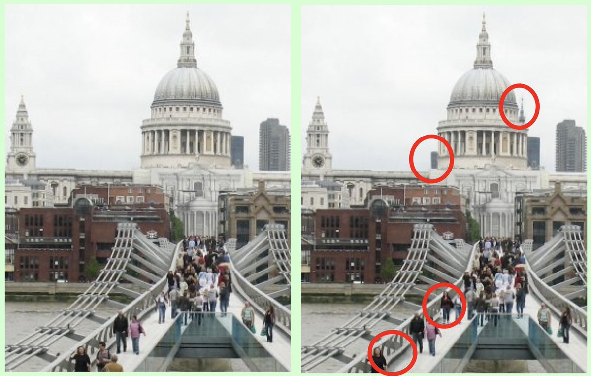
🧵4/16 Look at the scout. One time we missed an intracranial abscess that showed intracranial air only on the scout image, but the actual sinus CT images did not cover that intracranial area. #radres #MedEd #futureradres #radiology ajronline.org
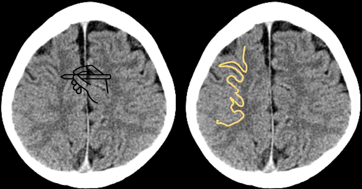
🧵5/16 Use this mental tool to assess for gray-white matter differentiation: Imagine a pen in your hand- can you confidently draw a line along the gray-white border? If not, consider the presence of loss of gray-white differentiation #radres #MedEd #radiology #futureradres
🧵 6/16 Assessing sulci. Look for 3 shades of gray: Gray matter-lightest shade (yellow), white matter-darker shade (blue), CSF -lowest shade (purple). If CSF shade is missing, consider sulcal effacement (from mass effect) or obscuration of CSF (from blood or infectious debris)

🧵7/16 Use these mental tools to assess for basilar cistern effacement: normal pentagon or square shape of the suprasellar cistern and the smiley face of the quadrigeminal plate cistern. If missing- worry about mass effect/herniation! #radres #MedEd #radiology #futureradres

🧵8/16 Types of bleeds. Epidural: Biconvex & crosses suture lines, Subdural: Crescentic, Subarachnoid: Replaces CSF & extends deep into sulci, Subpial: Follows the superficial outline of the brain & does not go deep into sulci. stritch.luc.edu
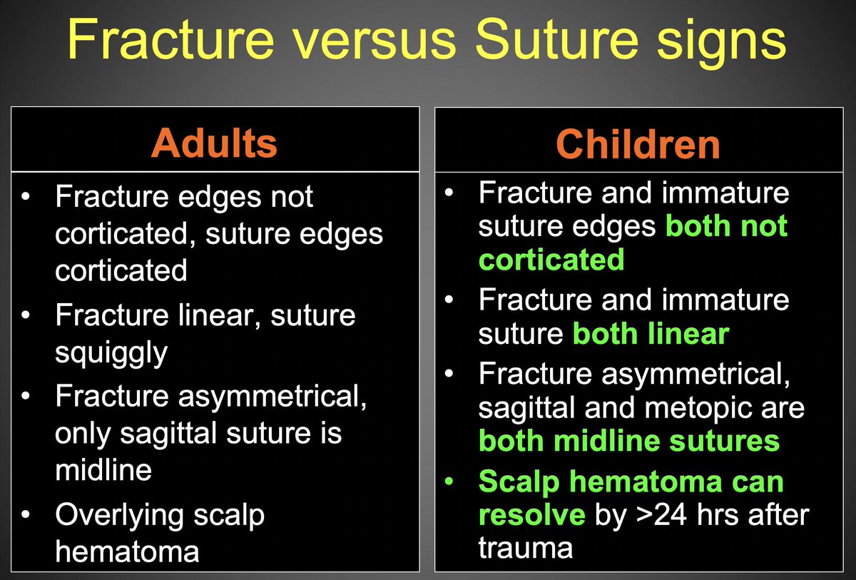
🧵9/16 Suture or fracture? This can be challenging as adult and pediatric skulls have some different features. Look for what you know- be familiar with normal skull sutures and accessory sutures to quickly identify fractures. #radres #MedEd #radiology #futureradres
🧵10/16 Life-long learning. We find what we look for. We look for what we know. While learning radiology appears to happen through the random appearance of cases on your worklist, there is a lot to be said for a systematic study approach to broaden your horizon.

🧵11/16 Importance of clinical info. Take the time review the chart and find the patient’s presenting symptoms, any working diagnoses that may be pursued, and any pertinent patient history. This will help you significantly in knowing what to look for. ncbi.nlm.nih.gov
🧵12/16 Stroke signs. There are sets of imaging signs that pertain to a specific clinical question, such a stroke, trauma, and shunt malfunction, to name just a few. Here is a great introductory resource for CT stroke imaging signs: radiologyassistant.nl #radres #MedEd
🧵13/16 Head trauma signs. There are sets of imaging signs that pertain to a specific clinical question, such a stroke, trauma, and shunt malfunction, to name just a few. Here is a great introductory resource for CT trauma imaging signs: radiopaedia.org #radres #MedEd
🧵14/16 Signs of shunt malfunction. There are sets of imaging signs that pertain to a specific clinical question, such a stroke, trauma, and shunt malfunction, to name just a few. Here is a great introductory resource for shunt imaging: www-sciencedirect-com.proxy.library.emory.edu
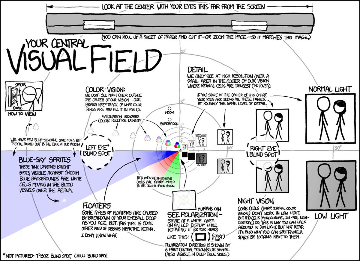
🧵15/16 Consider your center of vision. Dividing the PACS monitor into smaller windows can help putting more imaging information into the center of your visual field (rather than using the entire monitor and wander around with your eyes). Whatever you do- review ALL images.
🧵16/16 Last but not least: It really is a commitment to life-long learning, be aware that each advance from novice to expert carries its own risks. Take time to reflect on your journey. Be well and enjoy radiology!

Loading suggestions...