
Excited to share the latest preprint with @MikeAngeloLab! While imaging studies often focus on cell objects, images capture significant info outside of cells. Enter Pixie, a pipeline for the quantitative annotation of both pixel and cell level features🧵biorxiv.org
While segmentation algorithms (check out @NoahGreenwald's awesome work) can accurately identify cell boundaries in images, identifying cell phenotypes remains difficult, likely due to confounders such as plane of tissue sectioning, dense tissue, or irregularly shaped cells. [2/x]
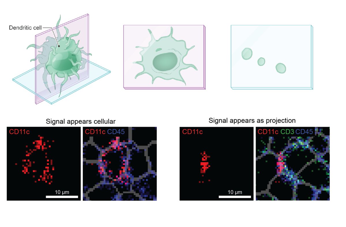
In tissues that contain large amounts of extracellular matrix or structural protein (such as this ex. of triple negative breast cancer), most pixels in the image are actually outside the cellular space. There is currently no straightforward way to quantify these features. [3/x]
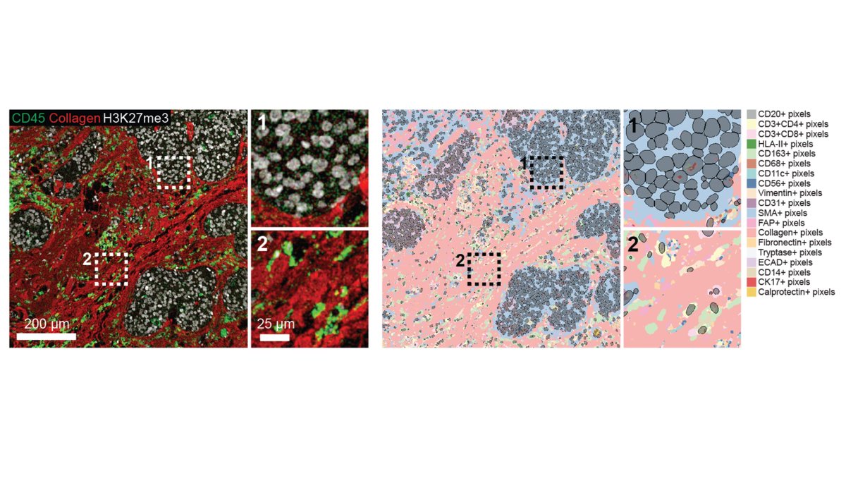
We developed Pixie, a pipeline that uses unsupervised clustering to assign pixel phenotypes (lymph node example shown). We perform extensive evaluation of pre-processing steps and parameter choices to optimize clustering, as well as comprehensively assess reproducibility. [4/x]
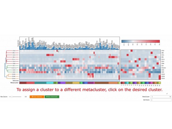
Unsupervised clustering methods rely on manual annotation to assign clusters to phenotypes. To balance automation with human curation, we created a simple GUI for easily making adjustments to clusters and annotations (left: 100 clusters, right: metaclusters of these 100). [5/x]
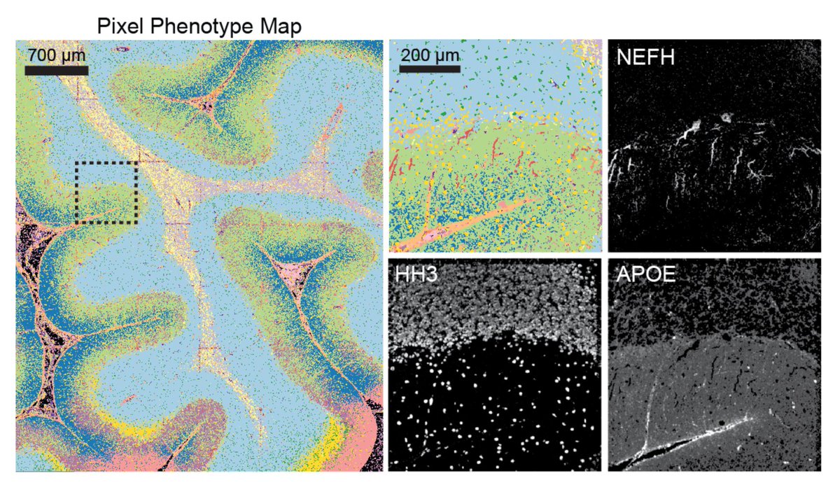
Due to abnormal shapes and complex spatial conformations of neuronal objects, cell segmentation is difficult in brain images. In beautiful MIBI images by @Dunja_Mrdjen, Pixie was able to map the full neuronal landscape, including neurons, vessels, astrocytes, and microglia. [7/x]
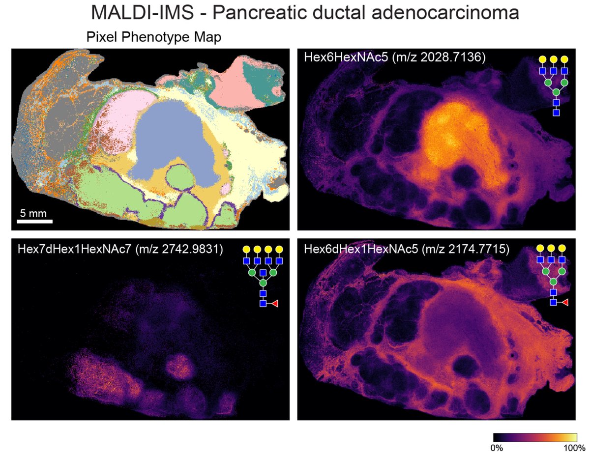
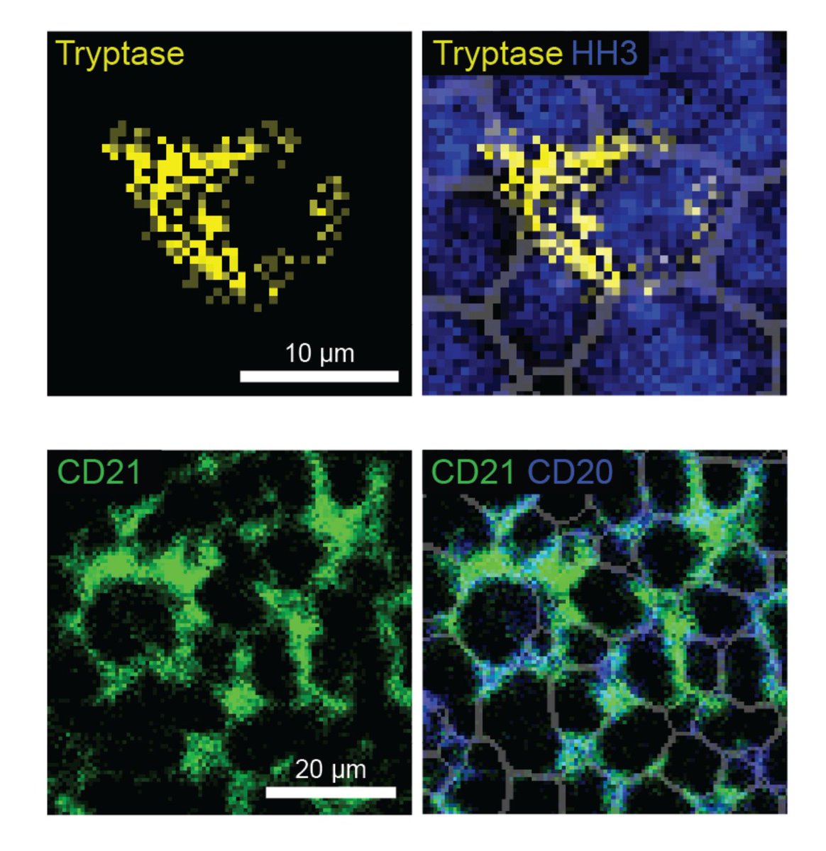
We show applications across other platforms, including fluorescence-based CODEX and label-free MALDI-IMS (used to map N-linked glycans). Even though these are completely different technologies that capture different molecules, Pixie was able to capture informative features. [8/x]
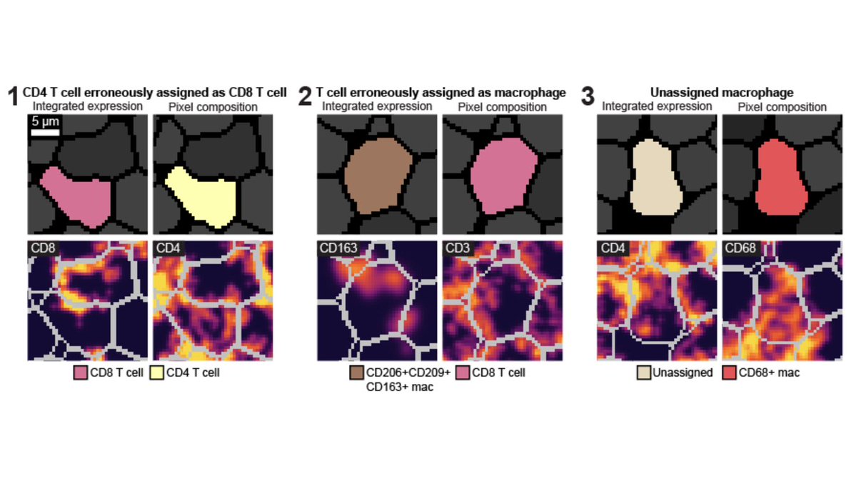
The current paradigm for assigning cell phenotypes is to cluster using integrated marker expression for each cell. However, this often results in poor cluster definition that requires a lot of manual work to adjust. This could be due to noisy pixels or abnormal shapes. [9/x]
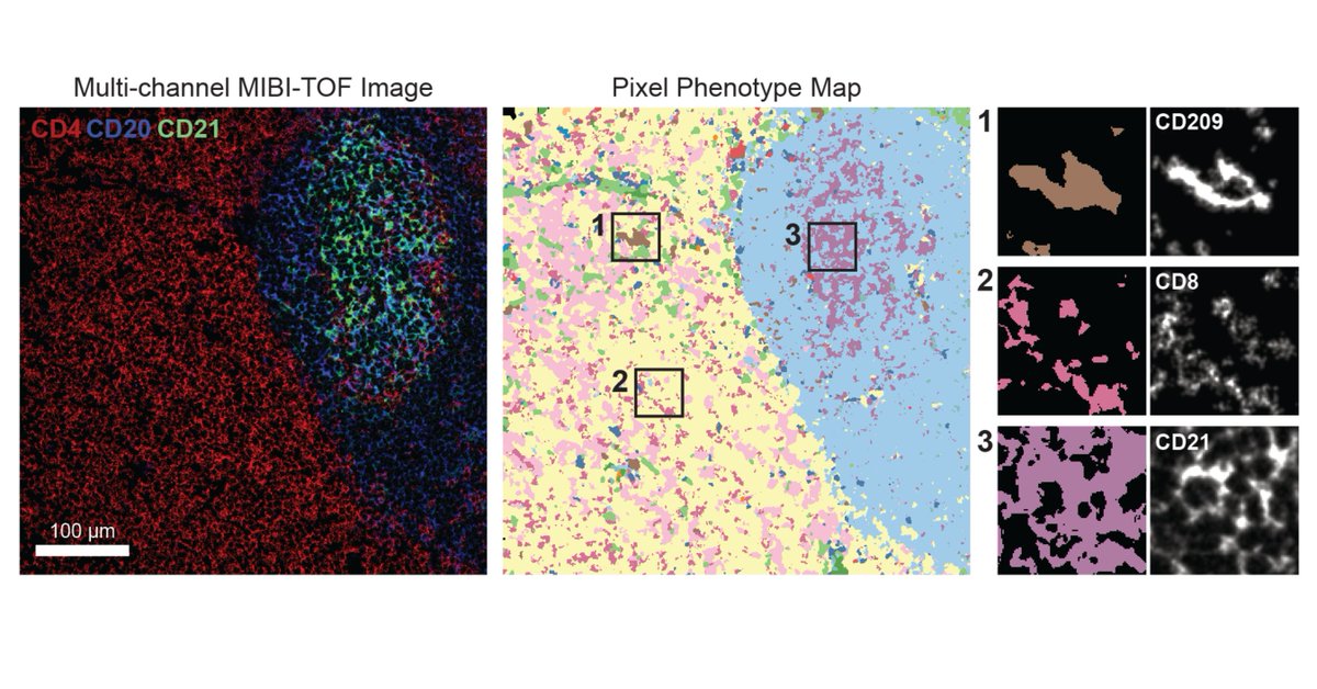
By using pixel features instead of integrated expression to perform cell clustering, Pixie results in better cluster definition and reproducibility. We show specific examples of where Pixie is advantageous to using integrated expression for cell phenotyping. [10/x]
Taken together, Pixie is a simple, scalable pipeline that can generate quantitative annotations of features both independently and in conjunction with segmentation. Pixie is available as user-friendly Jupyter notebooks at github.com. Try it out! [11/x]
Big thanks to all my co-authors, @NoahGreenwald, Alex Kong, @ErinMcCaffrey14, Ke Leow, and @Dunja_Mrdjen. Who knew a quick "Can you try this one quick analysis for me?" from @MikeAngeloLab would turn into a whole paper? [12/end]
Loading suggestions...