

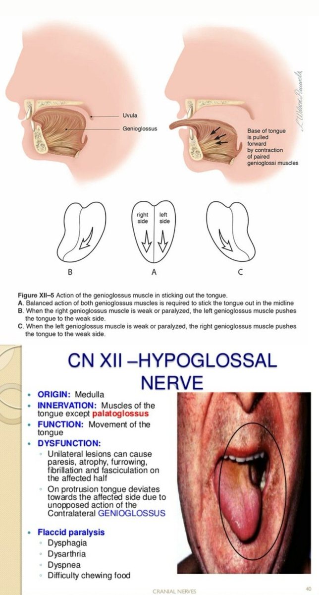
What cranial nerve palsy is most clearly illustrated in this image?
Left facial nerve
Left glossopharyngeal nerve
Left hypoglossal nerve
Right glossopharyngeal nerve
Right hypoglossal nerve
Left facial nerve
Left glossopharyngeal nerve
Left hypoglossal nerve
Right glossopharyngeal nerve
Right hypoglossal nerve

A 45-year-old woman
👉3 days after the acute onset of severe dysphagia, breathy dysphonia, and earache and pulsatile tinnitus in the left ear.
👉3 days after the acute onset of severe dysphagia, breathy dysphonia, and earache and pulsatile tinnitus in the left ear.
👉 medical history was unremarkable
👉 examination revealed
👉leftward deviation of the protruded tongue that was consistent with a lesion in cranial nerve XII
👉 examination revealed
👉leftward deviation of the protruded tongue that was consistent with a lesion in cranial nerve XII
👉hypernasal speech and rightward deviation of the soft palate on phonation
👉 consistent with lesions in cranial nerves IX and X
👉 paralyzed left vocal cord detected on laryngoscopic examination that was consistent with a lesion in cranial nerve X (see video).
👉 consistent with lesions in cranial nerves IX and X
👉 paralyzed left vocal cord detected on laryngoscopic examination that was consistent with a lesion in cranial nerve X (see video).

Axial MRI of the head and neck
👉 an extracranial dissection of the left internal carotid artery (see video)
👉 with delayed perfusion of the left hemisphere
👉 no evidence of ischemic stroke.
👉 an extracranial dissection of the left internal carotid artery (see video)
👉 with delayed perfusion of the left hemisphere
👉 no evidence of ischemic stroke.
👉 acute onset of cranial-nerve palsies accompanied by pain in the head, neck, or ear
👉 consider a diagnosis of internal-carotid-artery dissection
Most peripheral palsies are associated with cranial nerves IX through XII
👉 consider a diagnosis of internal-carotid-artery dissection
Most peripheral palsies are associated with cranial nerves IX through XII
👉 patient was treated conservatively
👉 showed complete neurologic recovery at a 6-month follow-up examination.
👉 showed complete neurologic recovery at a 6-month follow-up examination.
@jayukids These points are useful

Loading suggestions...