
Another set of cardiac #POCUS #anatomy illustrations. 🧵
#Nephpearls #FOAMed
Source: #fig1" target="_blank" rel="noopener" onclick="event.stopPropagation()">sciencedirect.com
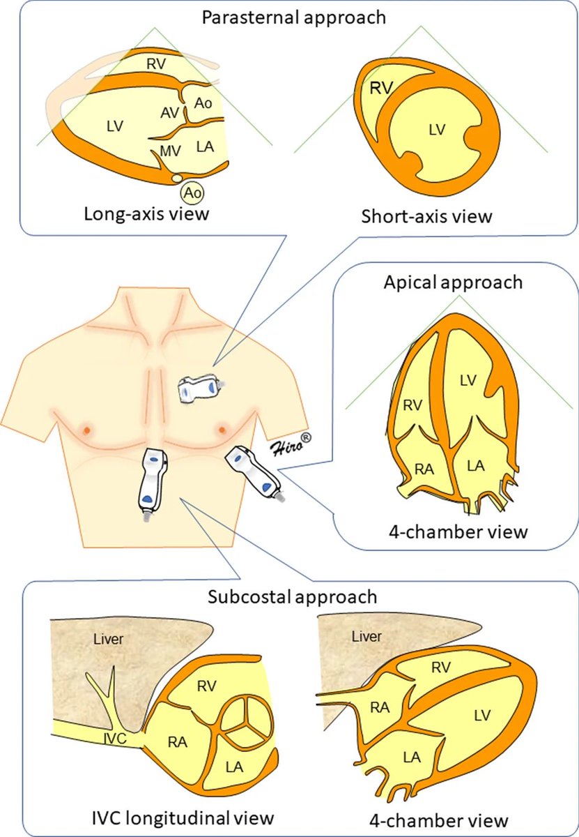
1⃣ Parasternal long axis
#Nephpearls #FOAMed
Source: #fig1" target="_blank" rel="noopener" onclick="event.stopPropagation()">sciencedirect.com
1⃣ Parasternal long axis

2⃣ Parasternal short axis aortic valve level
#POCUS
#POCUS

3⃣ Apical 4-chamber view #POCUS

4⃣ Subxiphoid 4-chamber view #POCUS


6⃣ Transesophageal #echofirst #Anatomy
As the beam is coming from the posterior aspect (esophagus), the image will be flipped compared to transthoracic echo.
As the beam is coming from the posterior aspect (esophagus), the image will be flipped compared to transthoracic echo.

Loading suggestions...