

I am pleased to announce that our newest review article (Drug-induced lung disease: a brief update for radiologists) has been published in the Diagnostic and Interventional Radiology.
@TLHM_MD @ctisus
dirjournal.org
@TLHM_MD @ctisus
dirjournal.org
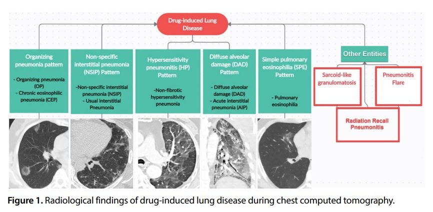
• Although Drug-induced lung disease (DILD) is difficult to distinguish clinically from other causes of diffuse pulmonary opacities, radiological findings play a key role.
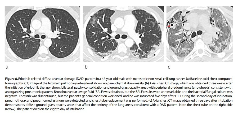
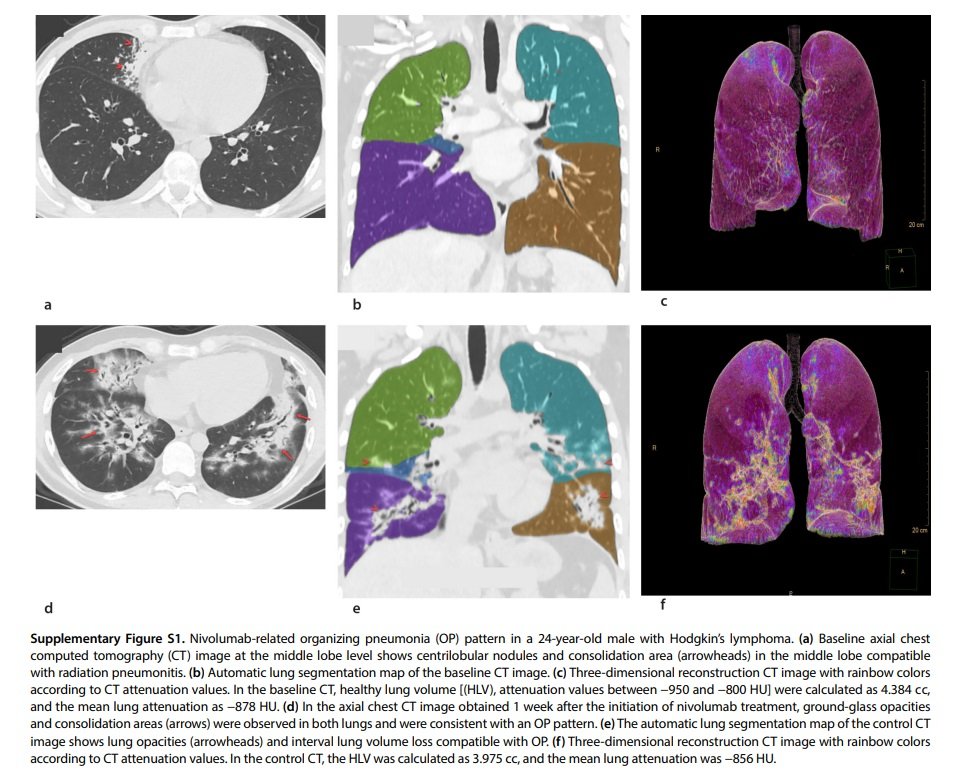
•The OP pattern is a form of acute lung injury and is the most common form of DILD
•The OP pattern is a form of acute lung injury and is the most common form of DILD
Although eosinophilic pneumonia (EP) can be present in similar imaging findings with an OP pattern, EP is characterized by peripheral band-like opacities and predominance in the upper lobes
The NSIP pattern is the second most
the common form of DILD and is associated
with a median of grade 1 toxic effects.
the common form of DILD and is associated
with a median of grade 1 toxic effects.
The DAD pattern is characterized by GGOs or dependent consolidation areas on imaging that usually affect the majority of, and sometimes, the entirety of the lung area.
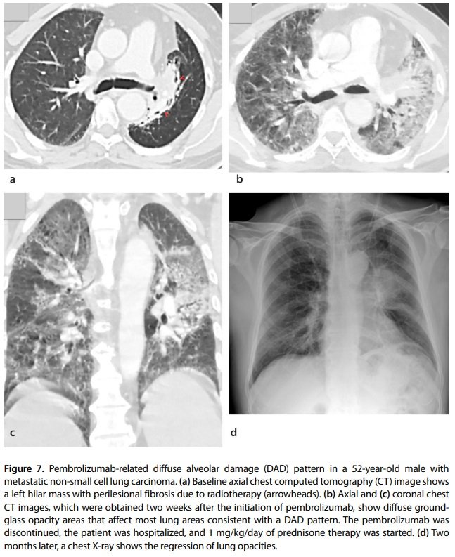
The “crazy paving” pattern characterized by interlobular septal thickening and intralobular lines can often be seen in the DAD pattern. In addition, other patterns, such as OP, can progress to DAD if not treated early.
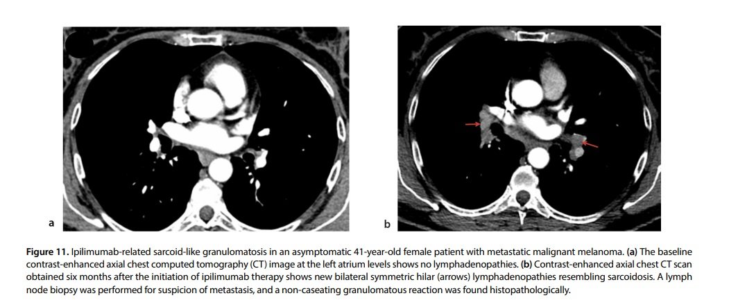
Sarcoid-like granulomatosis (SLG) is an atypical presentation of DILD and is usually associated with ICI therapy. SLG is characterized by histopathological and imaging features identical to sarcoidosis and includes enlarged lymph nodes and perilymphatic lung nodules.
Thin-slice chest CT is a valuable tool for a pattern-based assessment of DILD in the presence of appropriate medical history and clinical findings. Moreover, chest CT can reveal the severity of infiltrations in patients with DILD can be assessed visually or quantitatively.
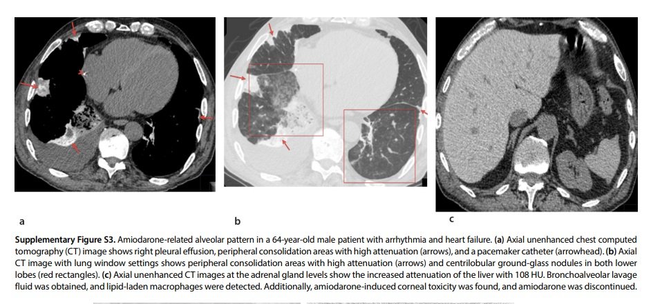
Amiodarone-related alveolar pattern in a 64-year-old male patient with arrhythmia and heart failure.
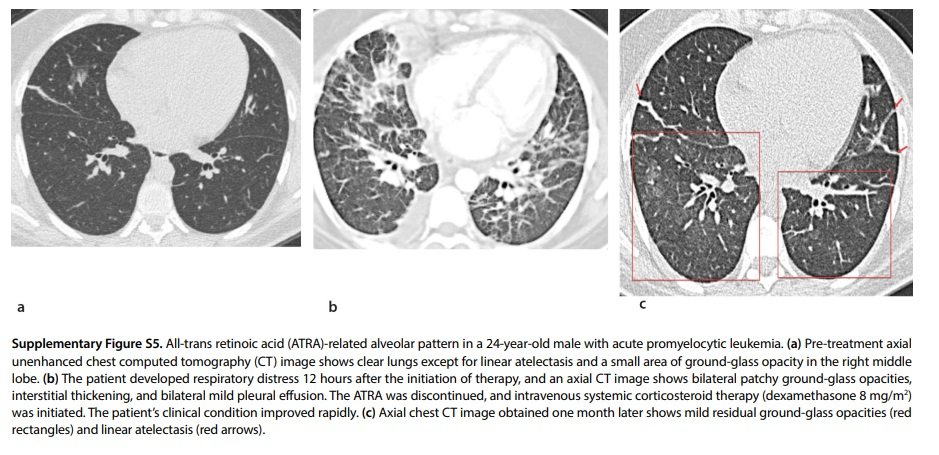
All-trans retinoic acid (ATRA)-related alveolar pattern in a 24-year-old male with acute promyelocytic leukemia.
Loading suggestions...