

Supportive imaging findings of idiopathic intracranial hypertension (pseudotumor cerebri)
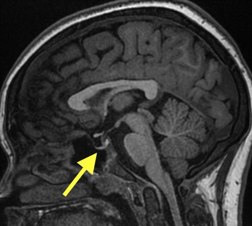
1️⃣Empty/partially empty sella
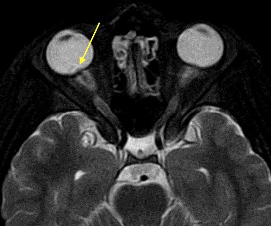
2️⃣Tortuous optic nerves with intraocular protrusion
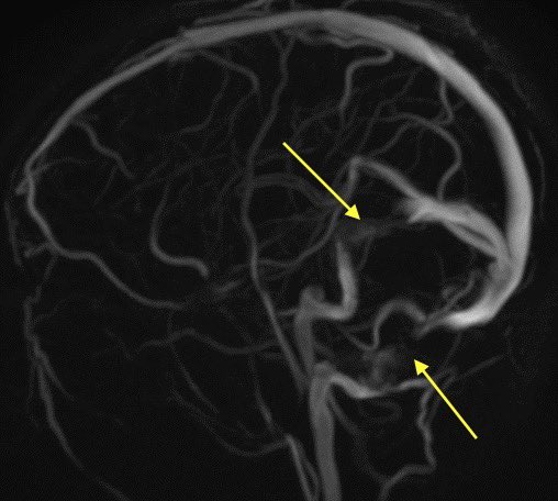
3️⃣Stenosis of transverse/sigmoid sinus junctions
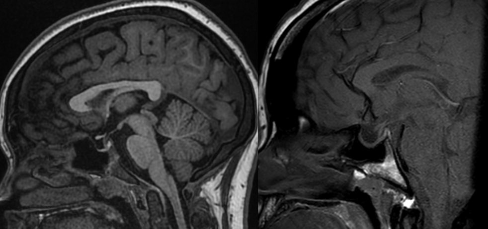
4️⃣Meningoencephaloceles
#Neurology #MedTwitter #radres
1️⃣Empty/partially empty sella
2️⃣Tortuous optic nerves with intraocular protrusion
3️⃣Stenosis of transverse/sigmoid sinus junctions
4️⃣Meningoencephaloceles
#Neurology #MedTwitter #radres

Presentation: classically, obese young and middle age women with headache and papilledema which may progress to vision loss
Etiology: unclear if due to increased intracranial volume (CSF, arterial pressure, venous volume, etc) or 2/2 decreased CSF/venous outflow
💡 Venous sinus stenosis may be a contributing cause or effect or increased pressure
💡 Venous sinus stenosis may be a contributing cause or effect or increased pressure
Imaging findings: All findings can be remembered if you understand that they are due to increased ICP
1️⃣Empty/partially empty sella: increased pressure results in forceful CSF pulsations which compresses the pituitary gland and empties the sella 🧠
1️⃣Empty/partially empty sella: increased pressure results in forceful CSF pulsations which compresses the pituitary gland and empties the sella 🧠

1️⃣Empty/partially empty sella, just remember this is also a normal incidental finding of aging as years and years of CSF pulsations can cause a flattened pituitary gland
Therefore, in the absence of other findings or appropriate history I do not mention empty sella after ~ 45yrs
Therefore, in the absence of other findings or appropriate history I do not mention empty sella after ~ 45yrs
2️⃣Tortuous optic nerves and intraocular protrusion: again, increased pressure leads to protrusion, tortuous nerves, and prominent CSF surrounding the nerves within the sheath

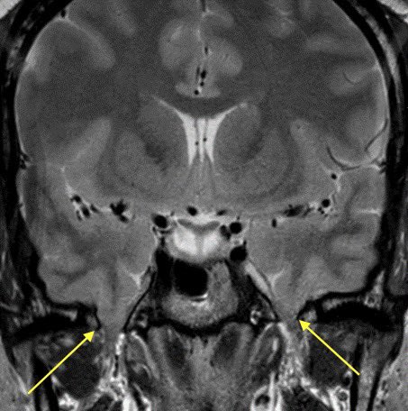
3️⃣Meningoencephaloceles: increased pressure pushes the meninges and brain through spaces they normally should not go. Here is an example of encephaloceles extending through the b/l foramen ovale 2/2 IIH
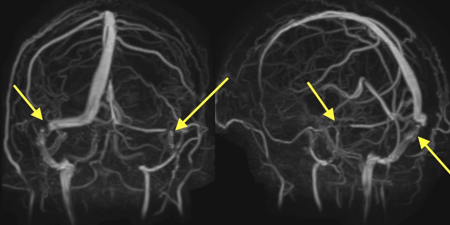
4️⃣Transverse/sigmoid sinus junction stenosis: stenosis may lead to the IIH or the increased pressure may lead to extrinsic compression of the sinuses, which comes first is unknown
Classically, this occurs at the junctions of transverse and sigmoid sinuses or lateral transverse
Classically, this occurs at the junctions of transverse and sigmoid sinuses or lateral transverse

Treatment:
1️⃣weight loss, acetazolamide (vaguely remember that from Med school)
2️⃣therapeutic LP or shunting
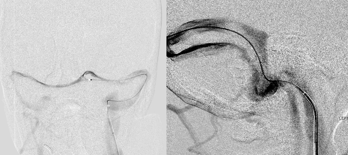
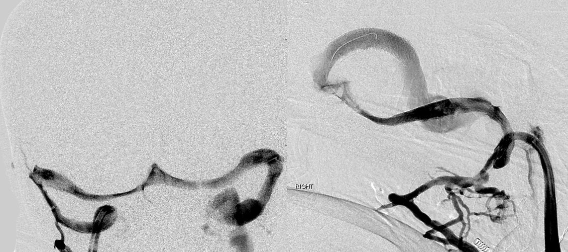
3️⃣Venous stent placement
Here is an example of an intracranial venogram pre and post stenting of the b/l Transverse/sigmoid sinus junctions
1️⃣weight loss, acetazolamide (vaguely remember that from Med school)
2️⃣therapeutic LP or shunting
3️⃣Venous stent placement
Here is an example of an intracranial venogram pre and post stenting of the b/l Transverse/sigmoid sinus junctions
Loading suggestions...