

Interesting case which I was wrong about prospectively. Let me share what I learned
Hx: 30 y/o M with severe occipital headache
Hx: 30 y/o M with severe occipital headache


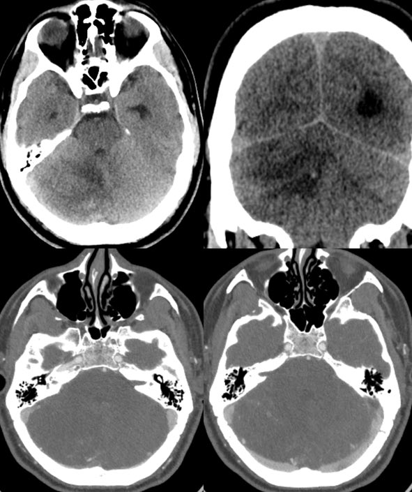
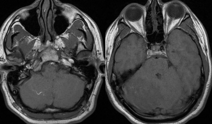
▶️Initial head CT demonstrates expansion and edema of the right cerebellar hemisphere. The overlying bones are unremarkable.
▶️CTA shows some peripheral increased vascularity but no thrombosis.
At this point the ddx is broad (tumor, infarct, infection, etc)
▶️CTA shows some peripheral increased vascularity but no thrombosis.
At this point the ddx is broad (tumor, infarct, infection, etc)
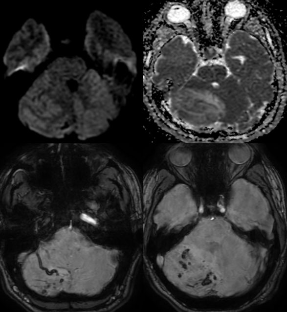
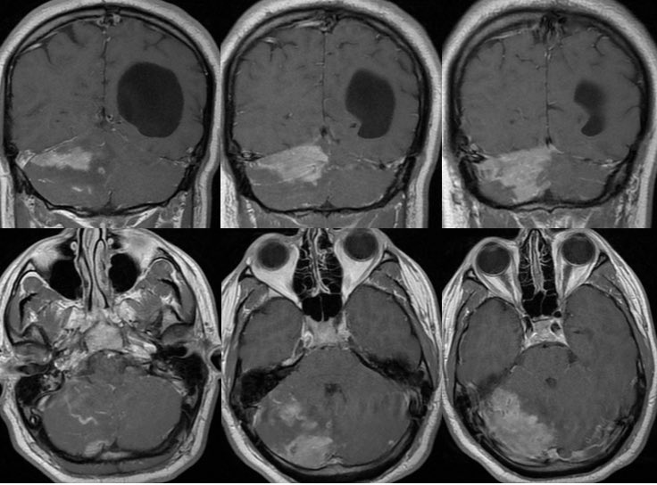
MRI: Enlarged or swollen right cerebellar hemisphere
T1: Hypointense with curvilinear high signal (slow flow in vessels)
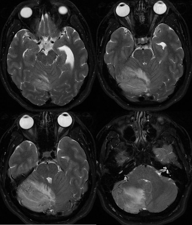
T2: multiple tortuous flow voids and extensive edema but PRESERVED FOLIA
T1C+: bizarre somewhat geographic enhancement of the parenchyma and leptomeninges
T1: Hypointense with curvilinear high signal (slow flow in vessels)
T2: multiple tortuous flow voids and extensive edema but PRESERVED FOLIA
T1C+: bizarre somewhat geographic enhancement of the parenchyma and leptomeninges
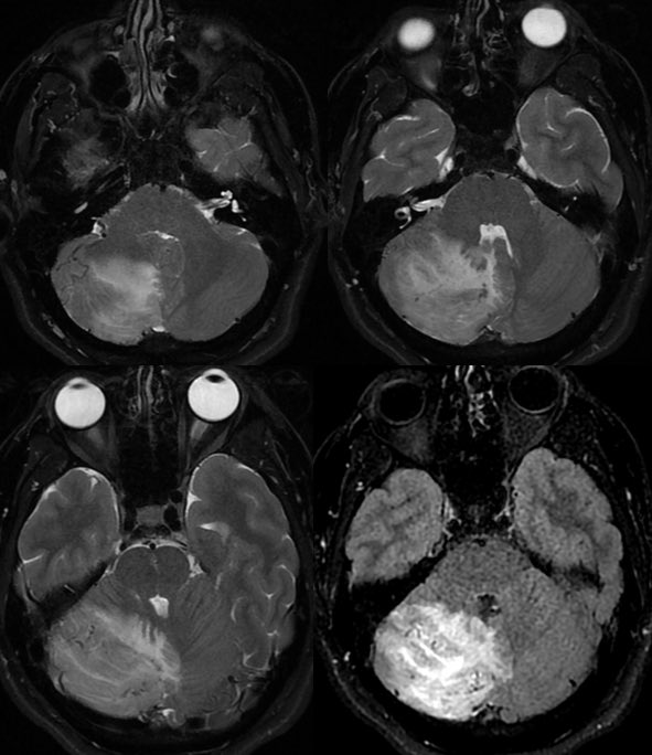


▶️DWI: No diffusion restriction
▶️SWI: patchy areas of hemorrhage and engorged vessels
▶️SWI: patchy areas of hemorrhage and engorged vessels

At this point:
Unilateral cerebellar mass effect those avid flow voids and enhancement may be due to tumor or ischemia
Unilateral cerebellar mass effect those avid flow voids and enhancement may be due to tumor or ischemia
Cerebellitis is a thought but the enhancement is too extensive within the parenchyma, the flow voids do not fit, and it would be strange to have such extensive unilateral disease
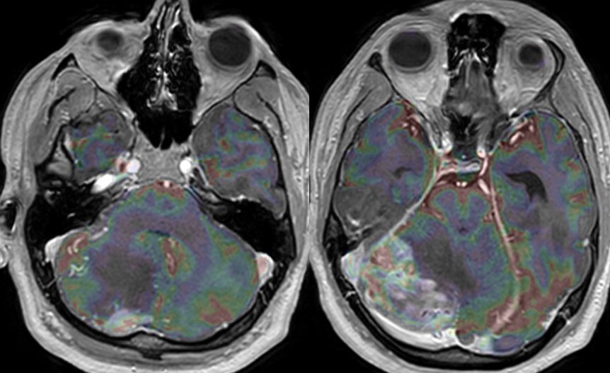
MR perfusion shows increased CBV within the enhancing components
MR perfusion shows increased CBV within the enhancing components
The lesion was surgically debulked
Final Dx: CNS vasculitis, unspecified
💡 IF THE FOLIA ARE PRESERVED, THINK ISCHEMIA (NOT TUMOR)
Infiltrating tumor would expand and distort the folia
Enhancement in this case was due to subacute infarct and inflammation
Final Dx: CNS vasculitis, unspecified
💡 IF THE FOLIA ARE PRESERVED, THINK ISCHEMIA (NOT TUMOR)
Infiltrating tumor would expand and distort the folia
Enhancement in this case was due to subacute infarct and inflammation
If you’re not sure, tumor vs vascular congestion/ischemia
💡 Consider short interval follow up
💡 DSA or ASL may have benefitted in this case as well 🧠
💡 Consider short interval follow up
💡 DSA or ASL may have benefitted in this case as well 🧠
Loading suggestions...