
[Thread 1/7]
Stay on topic : Male, 63yo, having chest pain for 30min without any effort
Initial EKG shows an inferior STEMI (OMI) + conduction impairment of the P wave, compatible with a type 2 AVB.
Vitals are ok
"Classic" initial ttt + pads pre-positionned on the patient.
Stay on topic : Male, 63yo, having chest pain for 30min without any effort
Initial EKG shows an inferior STEMI (OMI) + conduction impairment of the P wave, compatible with a type 2 AVB.
Vitals are ok
"Classic" initial ttt + pads pre-positionned on the patient.
![[Thread 1/7]
Stay on topic : Male, 63yo, having chest pain for 30min without any effort
Initial EKG...](https://pbs.twimg.com/media/FuoR3GbWYAEsaqd.jpg)
[2/7]
26min later, while in the elevator to the cath lab :
Some (multifocal, hardly visible on the EKG) PVCs starting to show, then NSVT.
26min later, while in the elevator to the cath lab :
Some (multifocal, hardly visible on the EKG) PVCs starting to show, then NSVT.
![[2/7]
26min later, while in the elevator to the cath lab :
Some (multifocal, hardly visible on the E...](https://pbs.twimg.com/media/FuoTHfIXgAAhuMJ.jpg)
[3/7]
3min later, just before being installed on the angio table, patient's rythm turns to FV.
Immediate shock x2, antero-lateral vector.
Neither vector change nor double def have been done, for ergonomic reasons.
3min later, just before being installed on the angio table, patient's rythm turns to FV.
Immediate shock x2, antero-lateral vector.
Neither vector change nor double def have been done, for ergonomic reasons.
![[3/7]
3min later, just before being installed on the angio table, patient's rythm turns to FV.
Immed...](https://pbs.twimg.com/media/FuoWg4cXwAAFG2t.jpg)
[4/7] After 3min of CPR, and after the 3rd shock, rythm changes to asystole.
Amiodarone had not yet been pushed.
We resumed the CPR, and started first mg of IV epinephrin.
Coro's senior decided not to perform angiography while CPR.
Amiodarone had not yet been pushed.
We resumed the CPR, and started first mg of IV epinephrin.
Coro's senior decided not to perform angiography while CPR.
![[4/7] After 3min of CPR, and after the 3rd shock, rythm changes to asystole.
Amiodarone had not yet...](https://pbs.twimg.com/media/FuoXT2YXgAIlze5.jpg)
[5/7]
ROSC 3min later, still showing inferior STEMI and PVCs on EKG.
NF 0 / LF 6min
The patient awakes. (GCS 15)
Coronary angiography is performed immediately after.
We are 35min from the initial EKG, and about 65min from the beginning of the chest pain.
ROSC 3min later, still showing inferior STEMI and PVCs on EKG.
NF 0 / LF 6min
The patient awakes. (GCS 15)
Coronary angiography is performed immediately after.
We are 35min from the initial EKG, and about 65min from the beginning of the chest pain.
![[5/7]
ROSC 3min later, still showing inferior STEMI and PVCs on EKG.
NF 0 / LF 6min
The patient awak...](https://pbs.twimg.com/media/FuoYNMsX0AAwsP1.jpg)
[6/7]
Unsurprisingly, an occluded RCA, featuring multilevel lesions.
Both the thrombo-aspiration and the angioplasty are successfull.
(The LCA is not unscathed, and will probably need a deferred intervention).
Unsurprisingly, an occluded RCA, featuring multilevel lesions.
Both the thrombo-aspiration and the angioplasty are successfull.
(The LCA is not unscathed, and will probably need a deferred intervention).
![[6/7]
Unsurprisingly, an occluded RCA, featuring multilevel lesions.
Both the thrombo-aspiration and...](https://pbs.twimg.com/media/FuoZEbQX0AE6HTI.jpg)
![[6/7]
Unsurprisingly, an occluded RCA, featuring multilevel lesions.
Both the thrombo-aspiration and...](https://pbs.twimg.com/media/FuoZFxQWYAQJdWq.jpg)
![[6/7]
Unsurprisingly, an occluded RCA, featuring multilevel lesions.
Both the thrombo-aspiration and...](https://pbs.twimg.com/media/FuoZGp6WwAIWjXA.jpg)
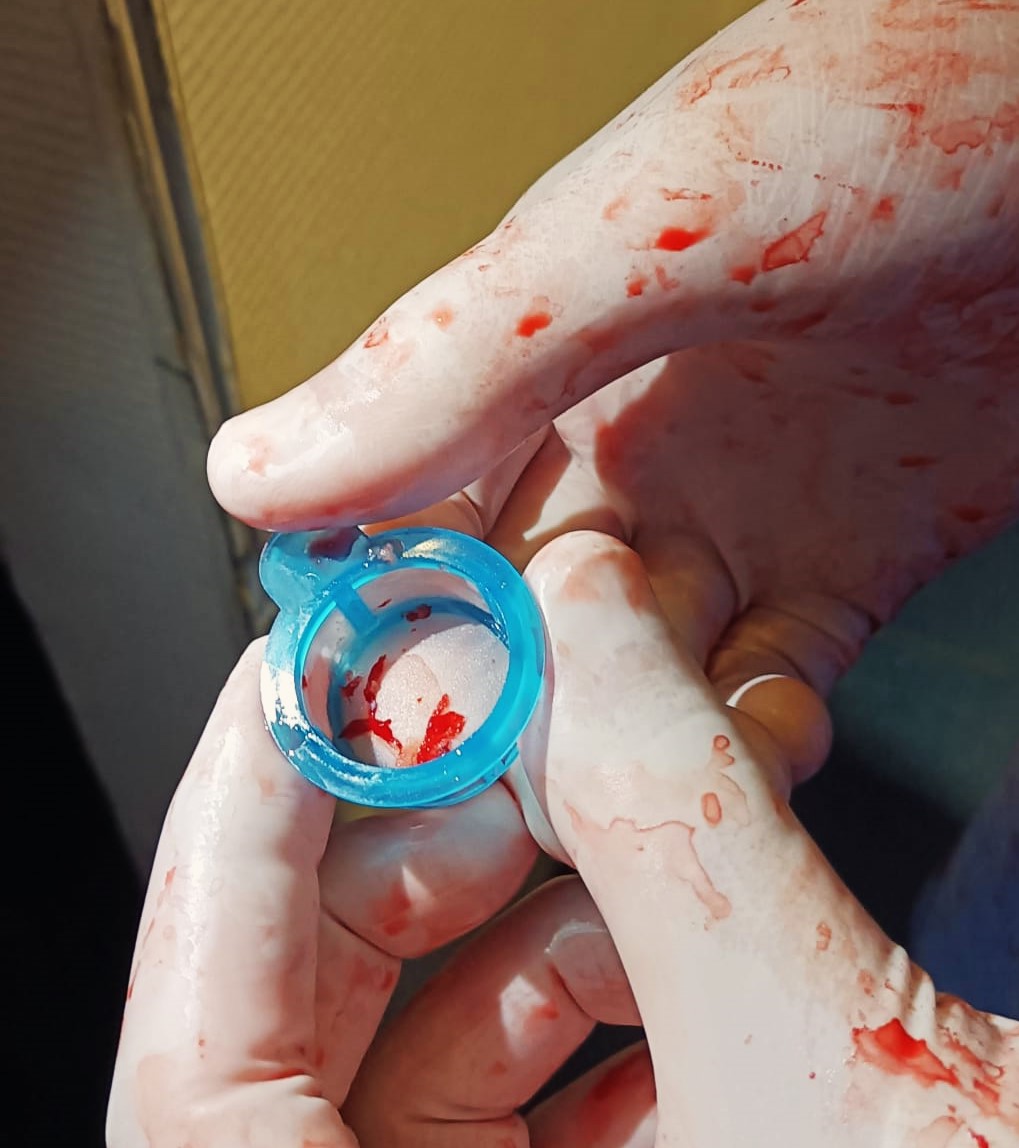
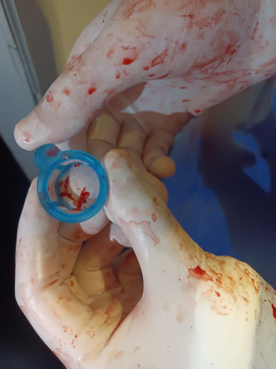
BONUS
The "guilty" clot responsible of the occlusion.
The "guilty" clot responsible of the occlusion.
Loading suggestions...