

Check out this new infographic🩻
One of my favorite blocks to perform - ultrasound-guided greater occipital nerve block. This graphic shows the important landmarks and ultrasound probe position to perform this block.🧵
One of my favorite blocks to perform - ultrasound-guided greater occipital nerve block. This graphic shows the important landmarks and ultrasound probe position to perform this block.🧵
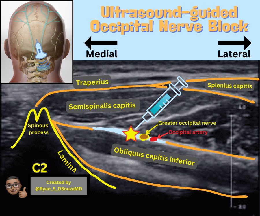
1/First start midline at the occiput and scan down until you see the bifid spinous process of C2. Then scan laterally to the side you are performing the block.
2/Optimize the image so that you see the bony dropoff of the C2 lamina as well as the muscle layers, especially the semispinalis capitis and the obliquus capitis inferior. The greater occipital nerve lies between these two muscles.
3/Don’t forget to use the Doppler view to scan for any important blood vessels near the vicinity of the block including the occipital artery. My needle trajectory is usually medial to lateral.
Loading suggestions...