

1/Having trouble remembering what to look for on scans for Alzheimer’s dementia?
Want the key findings to stay in your memory?
In honor of recent Alzheimer’s day, here's an easy way to semi-quantitate hippocampal volume for the evaluation of Alzheimer’s Dementia (AD)!
Want the key findings to stay in your memory?
In honor of recent Alzheimer’s day, here's an easy way to semi-quantitate hippocampal volume for the evaluation of Alzheimer’s Dementia (AD)!
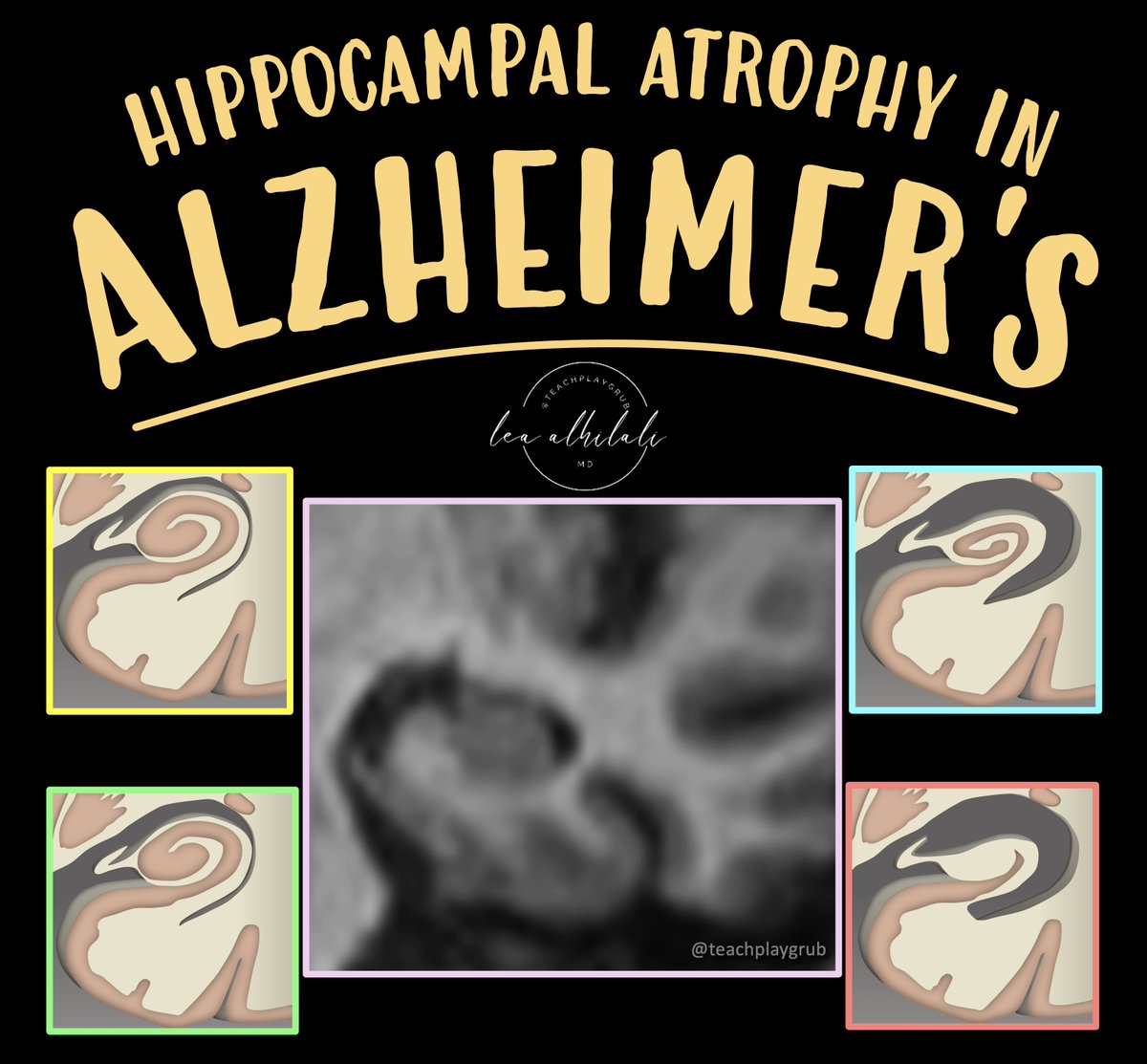
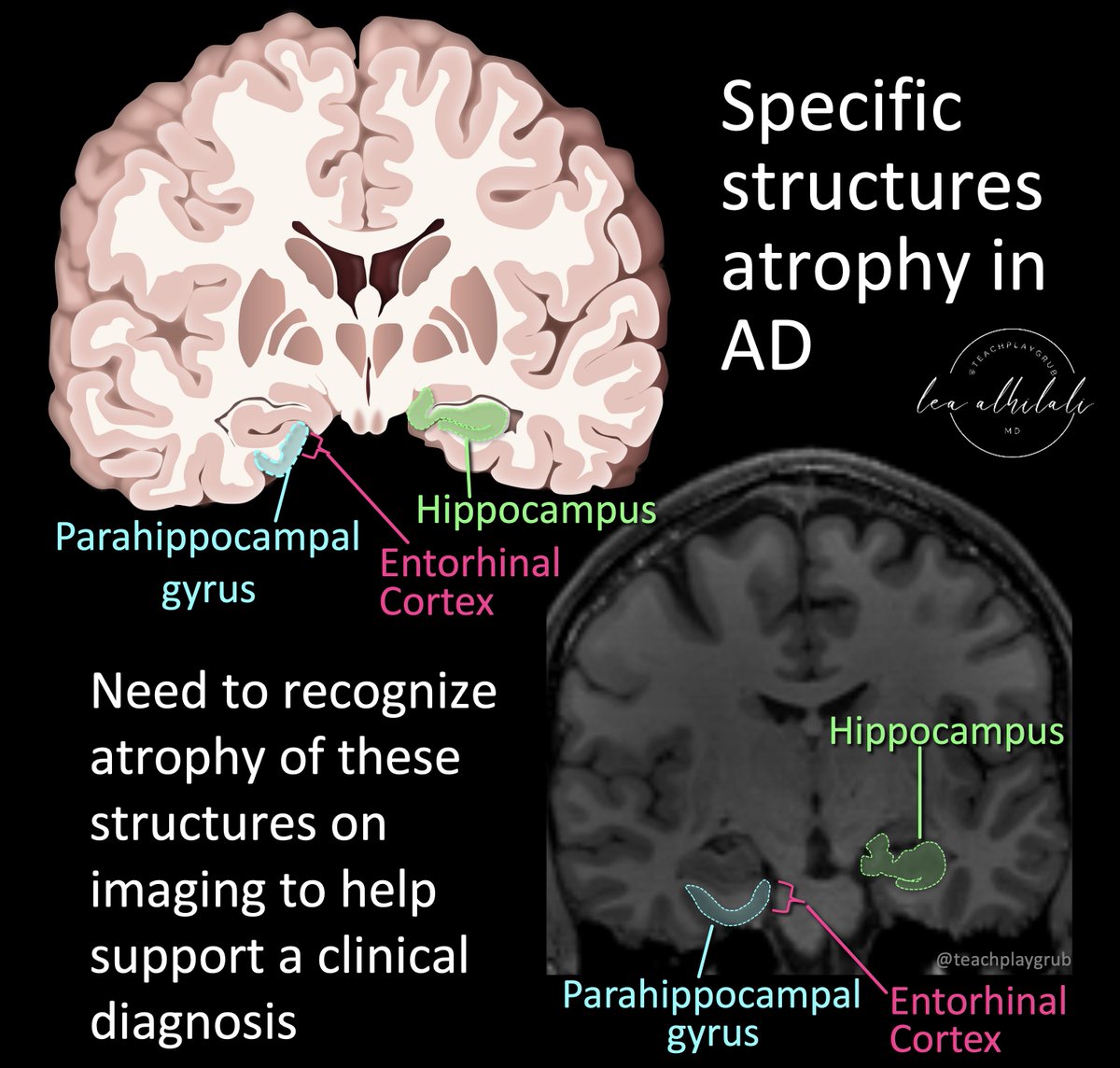
2/Specific patterns of atrophy are associated w/Alzheimer’s disease (AD).
Atrophy often involves the hippocampus & the anterior/medial portion of the adjacent parahippocampal gyrus, called the entorhinal cortex
This is where one should look for volume loss on imaging, but how?
Atrophy often involves the hippocampus & the anterior/medial portion of the adjacent parahippocampal gyrus, called the entorhinal cortex
This is where one should look for volume loss on imaging, but how?
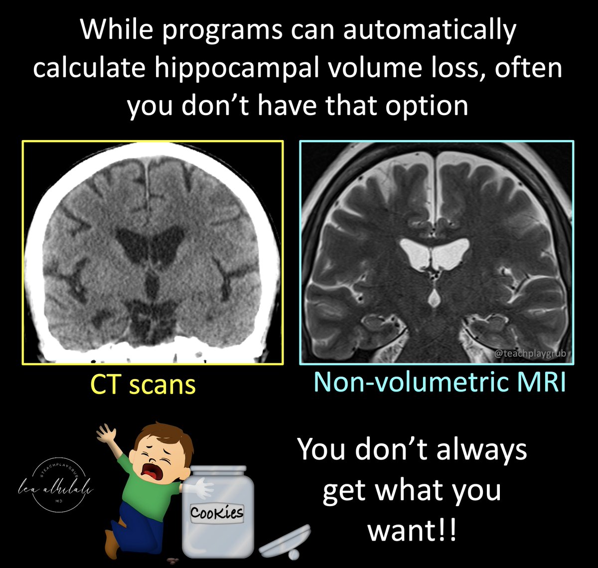
3/There are automated programs that can quantify hippocampal volume loss.
However, these can only be used on studies with volumetric imaging
But often times you have studies like CTs or routine MRIs that you would like to evaluate for AD.
Here, automated analysis cannot help you.
However, these can only be used on studies with volumetric imaging
But often times you have studies like CTs or routine MRIs that you would like to evaluate for AD.
Here, automated analysis cannot help you.
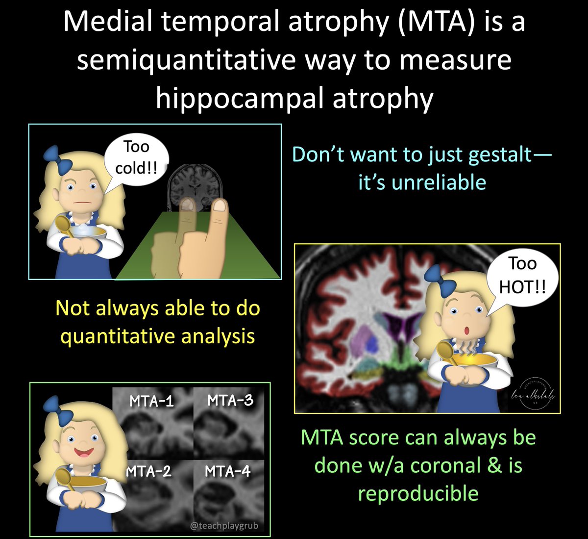
4/You don't want to just gestalt--it’s inaccurate & not reproducible.
But you often can't do automated analysis
Maybe the answer is somewhere in between--a very reproducible semiquantitative score, the medial temporal atrophy score
But you often can't do automated analysis
Maybe the answer is somewhere in between--a very reproducible semiquantitative score, the medial temporal atrophy score
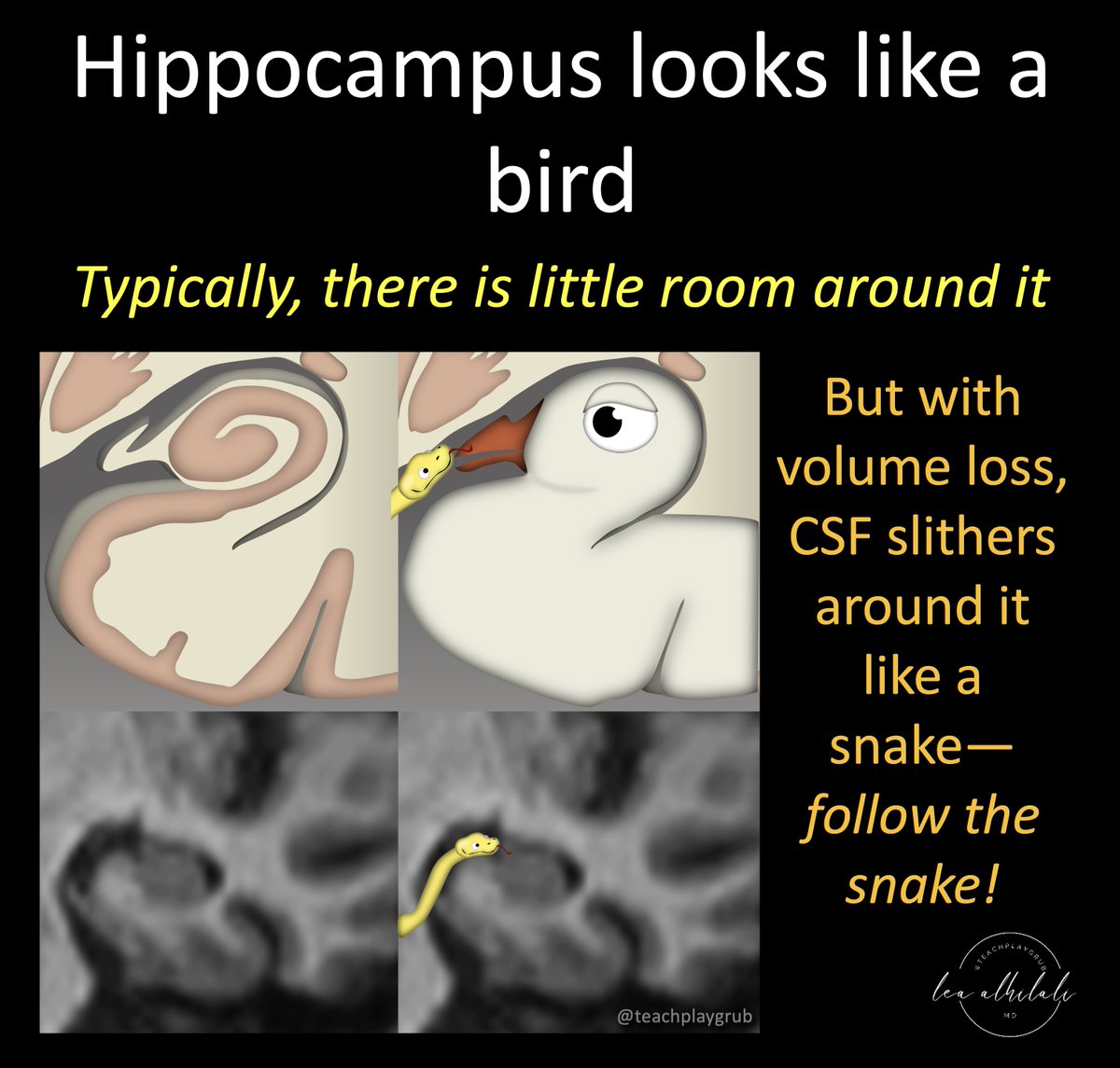
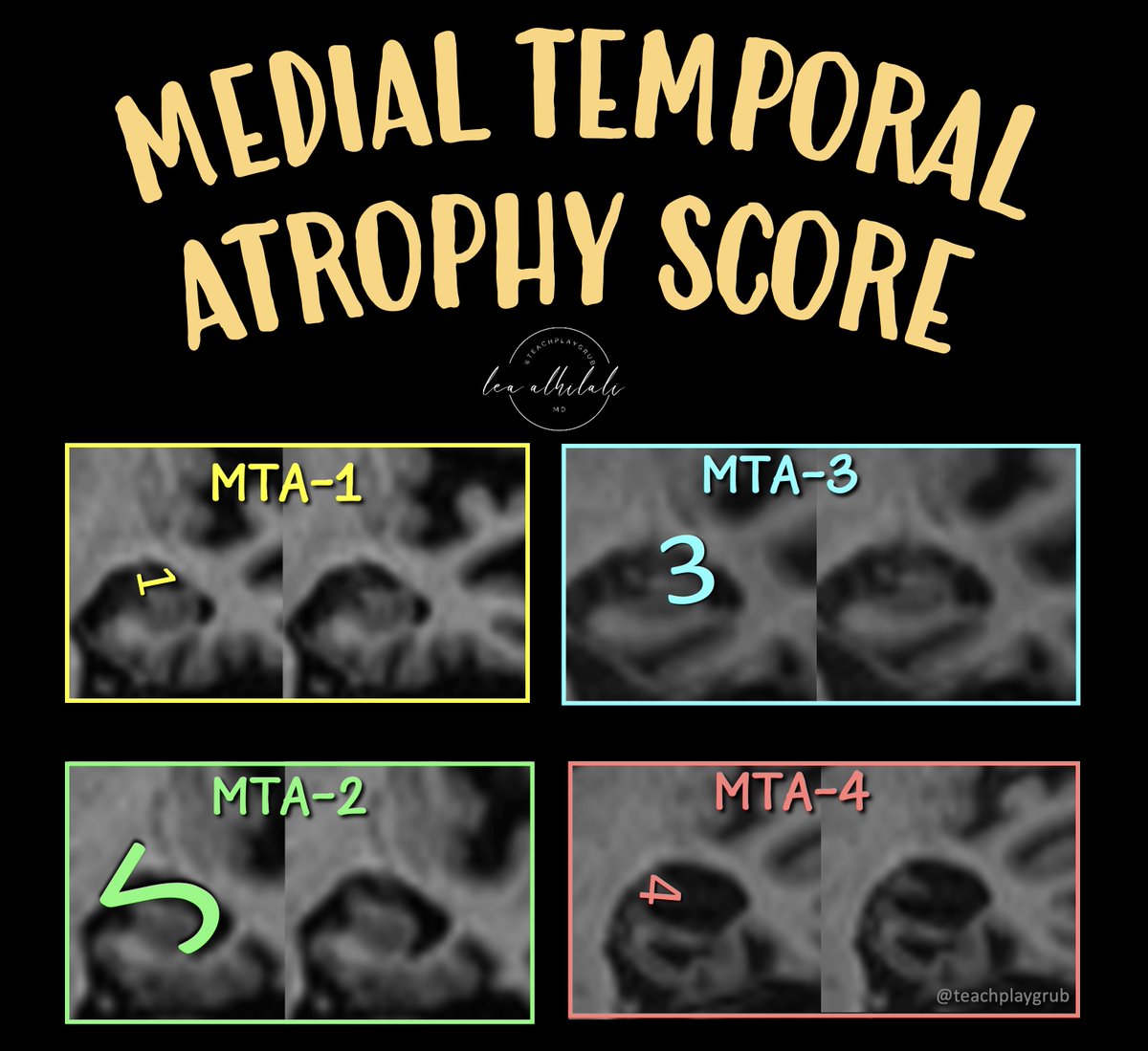
5/On coronal images, the hippocampus looks like a bird.
Typically, there is very little room around the bird’s head.
But w/volume loss, CSF slithers around like a snake.
MTA score grades how far the snake has gone towards eating the bird!
Typically, there is very little room around the bird’s head.
But w/volume loss, CSF slithers around like a snake.
MTA score grades how far the snake has gone towards eating the bird!
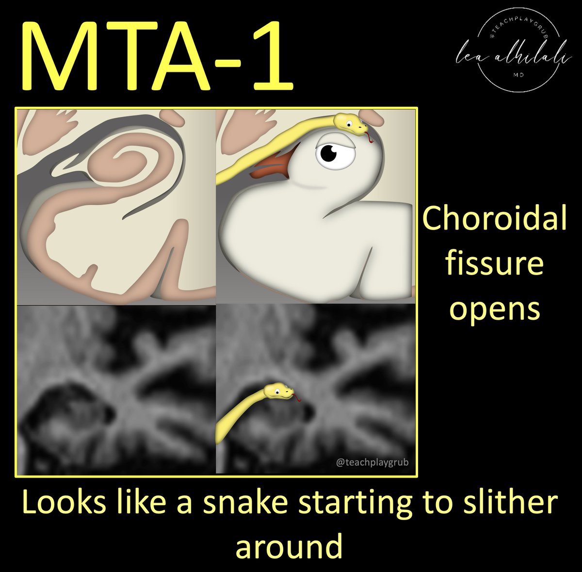
6/MTA-1 is the first step a snake takes towards eating the bird.
Here the choroidal fissure has slightly widened.
So the snake has begun to slither around the head of the bird.
Here the choroidal fissure has slightly widened.
So the snake has begun to slither around the head of the bird.
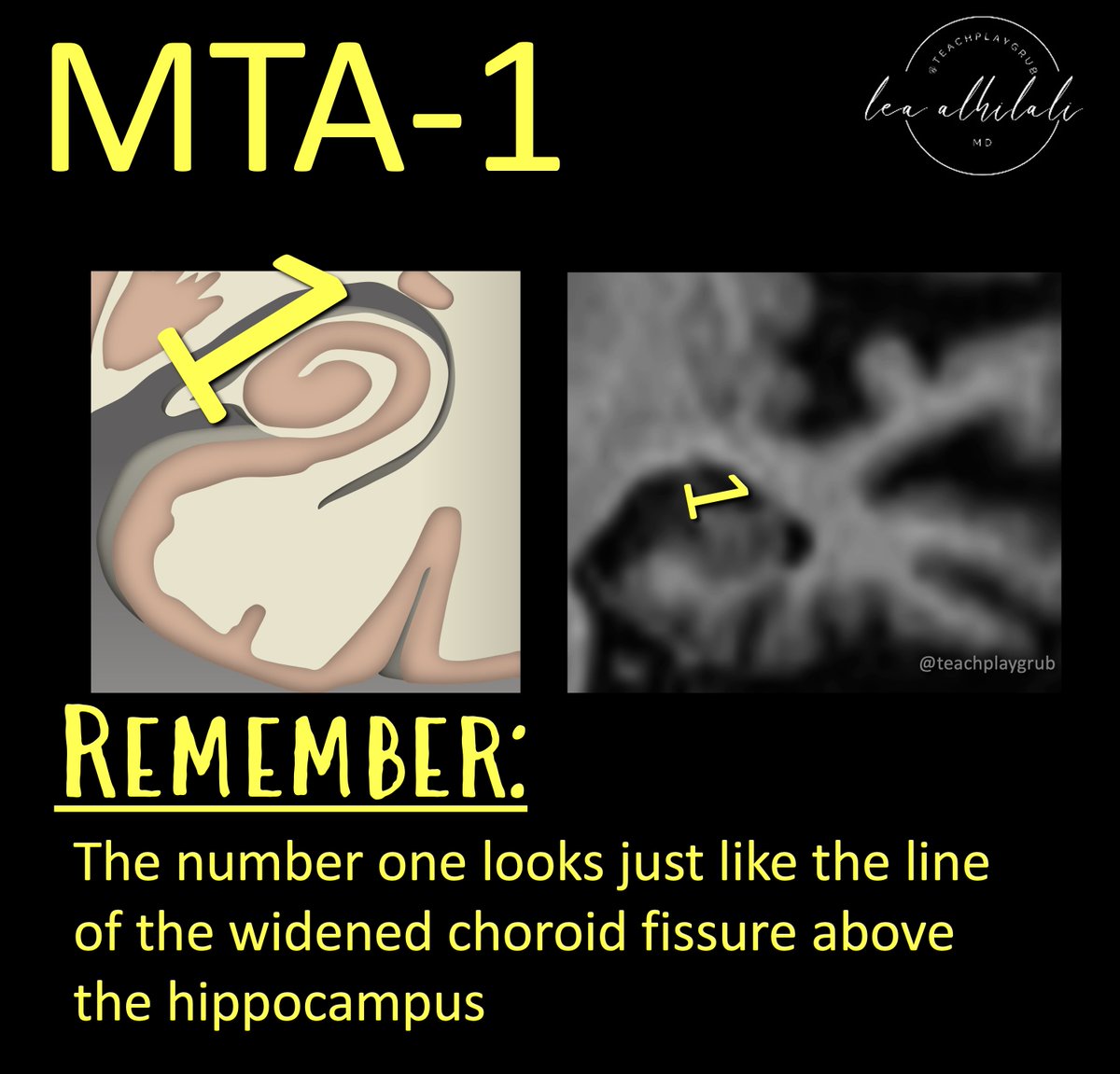
7/Remember this bc the number one looks flat like a slightly opened choroidal fissure!
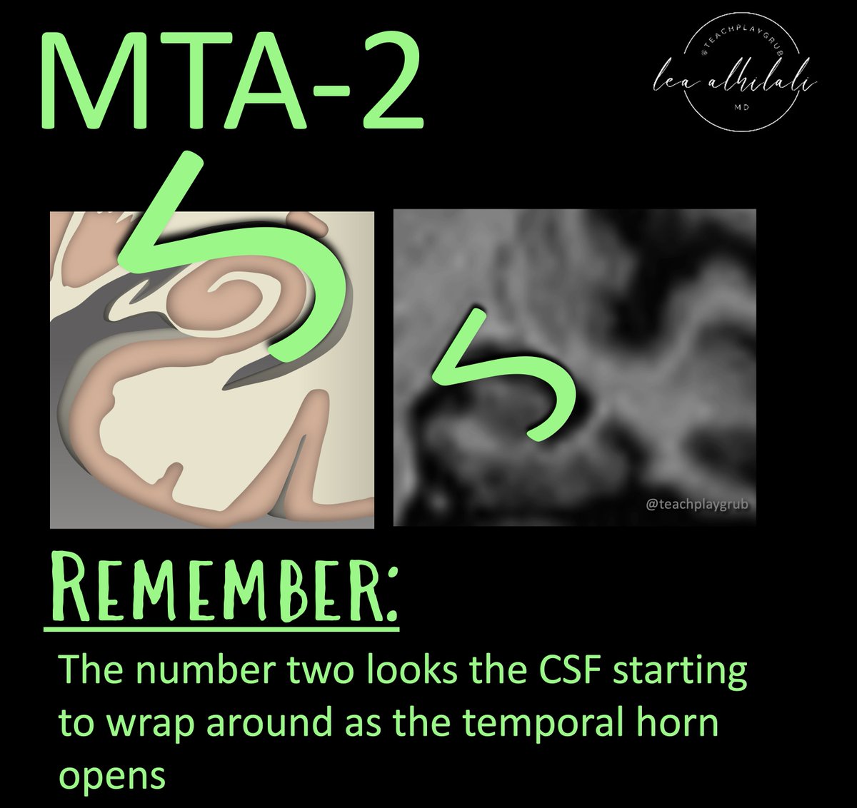
8/For MTA-2, the temporal horn also opens.
Now the snake has take the next step encircle the head of it’s bird victim.
Now the snake has take the next step encircle the head of it’s bird victim.
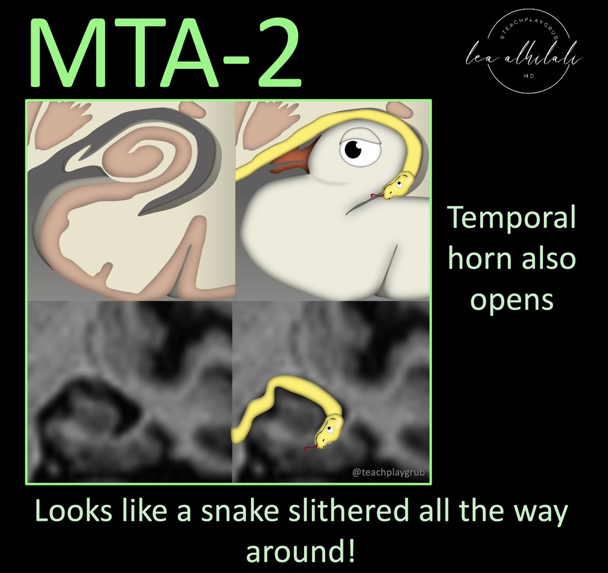
9/Remember this because the number two look like the CSF curving around as the temporal horn opens!
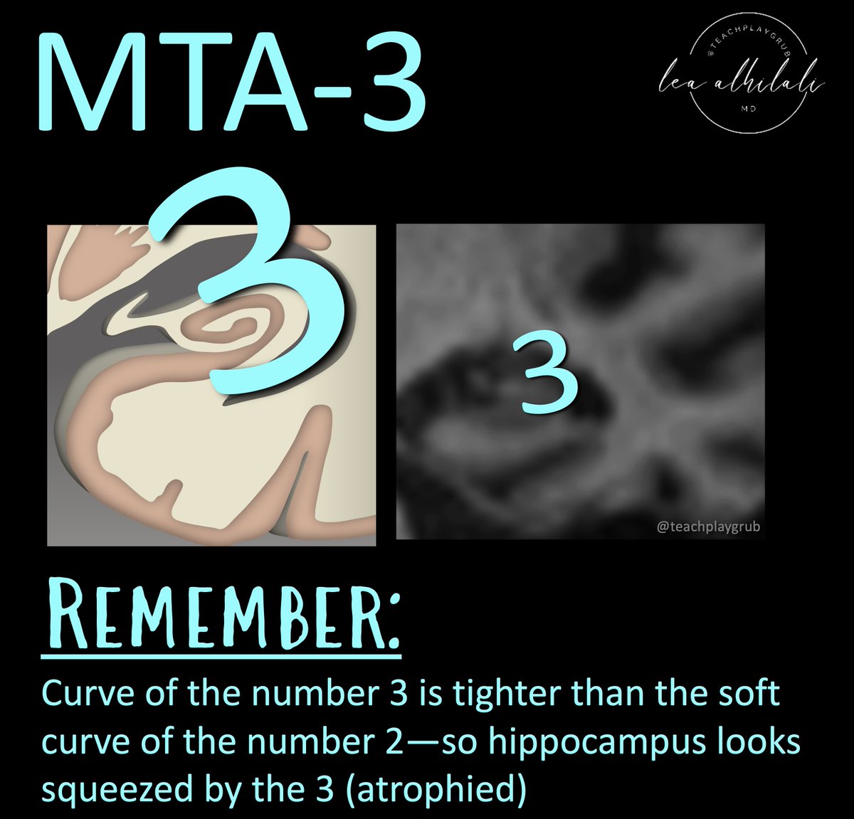
10/In MTA-3, the hippocampus shows moderate volume loss—so it looks like the head of the bird has been compressed.
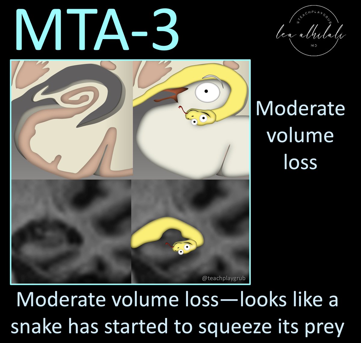
11/Remember this because the number three has a tight curve, so it looks like it is crushing the hippocampus & it looks small (atrophied)
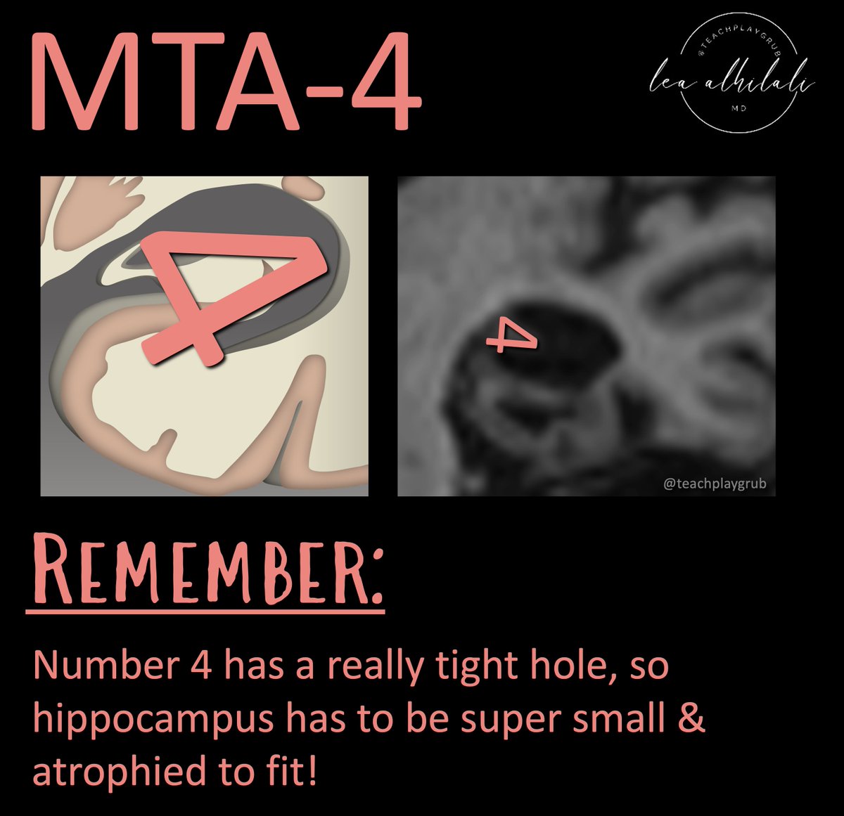
12/For MTA-4, the hippocampal volume loss is profound, so it looks like the head of bird has been crushed by the snake.
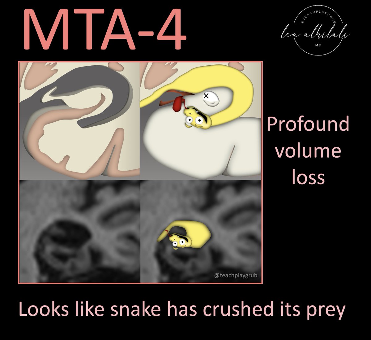
13/Remember this because the number four has only a tiny hole in it, so the hippocampus has to be super tiny & atrophied to fit inside!
14/Hopefully, this has made this Alzheimer dementia scoring system unforgettable!
Commit this rating system to memory so you’ll never forget what you need to say for AD!
Commit this rating system to memory so you’ll never forget what you need to say for AD!
Loading suggestions...