

1/
One of the BIGGEST #POCUS misconceptions is that B lines 🟰 pulmonary edema 🤦♀️
Did you know that there is actually a wide DDx for B lines?
Check out the latest @ubcimpocus video about #LungUltrasound for interstitial syndromes 👀; or read on for the highlights 👇 🧵
youtube.com
One of the BIGGEST #POCUS misconceptions is that B lines 🟰 pulmonary edema 🤦♀️
Did you know that there is actually a wide DDx for B lines?
Check out the latest @ubcimpocus video about #LungUltrasound for interstitial syndromes 👀; or read on for the highlights 👇 🧵
youtube.com
2/
Remember that B lines are generated because of something filling the (normally aerated) interstitium and interlobular septae.
Fluid can of course fill these spaces; but so can other things!
Pus, cells, fibrosis, blood - anything that can give you an interstitial pattern on a chest X ray can give you B lines on ultrasound.
3/
Broadly speaking, we can divide B lines into 2 categories. B lines arising from:
- Cardiogenic pulmonary edema 🫀
vs
- Infectious/inflammatory pathology 🦠
The latter includes things like atypical or viral pneumonias, ARDS, interstitial lung disease, lymphangitic carcinomatosis, etc.
Broadly speaking, we can divide B lines into 2 categories. B lines arising from:
- Cardiogenic pulmonary edema 🫀
vs
- Infectious/inflammatory pathology 🦠
The latter includes things like atypical or viral pneumonias, ARDS, interstitial lung disease, lymphangitic carcinomatosis, etc.
4/
And there are several ultrasound features that we can use to tease apart these two types.
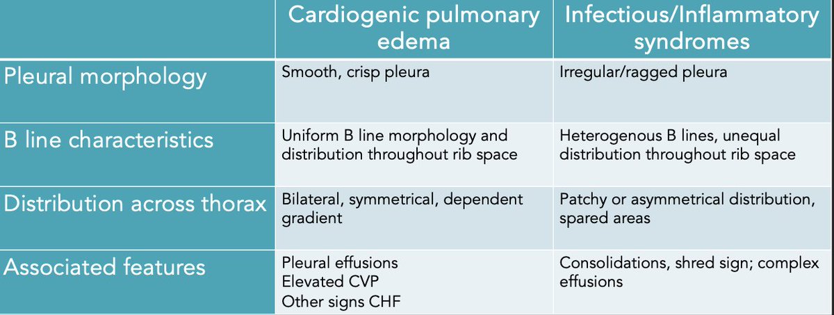
First: we can look at the pleural morphology.
B lines arising from a smooth, crisp, uniform pleural line are more likely to be from cardiogenic pulmonary edema. x.com
And there are several ultrasound features that we can use to tease apart these two types.
First: we can look at the pleural morphology.
B lines arising from a smooth, crisp, uniform pleural line are more likely to be from cardiogenic pulmonary edema. x.com

5/
In contrast, B lines arising from an irregular, ragged, interrupted pleural line are more likely to be from infectious/inflammatory pathologies.
(If you want to know WHY this happens - check out the video posted above!👆)
Note that it is key here for you as the scanner to use proper #LUS technique and employ fanning to ensure that your beam is perpendicular to the pleura.

6/
Next, we can look at the distribution of B lines across the interspace.
Pleural fluid fills interlobular septae in a predictable and regular way; which generates B lines that are evenly spaced throughout the whole interspace. x.com
Next, we can look at the distribution of B lines across the interspace.
Pleural fluid fills interlobular septae in a predictable and regular way; which generates B lines that are evenly spaced throughout the whole interspace. x.com

7/
In contrast, with infectious/inflammatory pathologies, you will see B lines which are NOT homogenous throughout the interspace.
You may see all the B lines arising from a single point on the pleura; or even A lines throughout part of the interspace. x.com
In contrast, with infectious/inflammatory pathologies, you will see B lines which are NOT homogenous throughout the interspace.
You may see all the B lines arising from a single point on the pleura; or even A lines throughout part of the interspace. x.com

8/
Next, we can look at the distribution of B lines across the thorax.
Note that this is why we always have to scan representative areas of BOTH sides of the chest - you cannot just take a single clip and call it pulmonary edema‼️
B lines arising from pulmonary edema should be bilateral, symmetrical, and in a dependent gradient (ie. most prominent at the bases).
B lines from infectious/inflammatory pathologies, on the other hand, are often asymmetrical, non-gravitational, and may display skipped or spared areas.

9/
Finally, we can look at other associated pathology.
In heart failure, for example, we will often see other features such as bilateral simple pleural effusions; plus cardiac findings supportive of CHF.
In inflammatory/infectious causes, you may see things like unilateral consolidations, dynamic air bronchograms, shred signs, complex effusions, etc.

10/
Of course - it goes without saying! - that all of these ultrasound findings should be incorporated with the rest of the clinical picture to help you reach a decision 🩺 x.com
Of course - it goes without saying! - that all of these ultrasound findings should be incorporated with the rest of the clinical picture to help you reach a decision 🩺 x.com
11/
Thanks for reading! 🙏
For more on #LungUltrasound, including image acquisition and common pitfalls - check out the rest of the videos on the ubcimpocus site ✨
Drop you #LUS pearls here! #MedTwitter #Tweetorial #POCUS #LungUltrasound
ubcimpocus.com
Thanks for reading! 🙏
For more on #LungUltrasound, including image acquisition and common pitfalls - check out the rest of the videos on the ubcimpocus site ✨
Drop you #LUS pearls here! #MedTwitter #Tweetorial #POCUS #LungUltrasound
ubcimpocus.com

Ping some fellow #POCUS nerds - @G2Disrupt @ria_dancel @DrGalenMD @ross_prager @KiranRikhraj @shanearish @BuddingSprout @HoosierPocus
Loading suggestions...