
🧵 Must-Know Signs in Neuroradiology: A Critical Thread for Clinicians & Neurosurgeons
Recognizing key radiological signs in neuroradiology is crucial for accurate diagnosis and management.
The essential "must-know" imaging signs every neurologist, neurosurgeon, and radiologist should recognize. 🧠🔍
👇
Recognizing key radiological signs in neuroradiology is crucial for accurate diagnosis and management.
The essential "must-know" imaging signs every neurologist, neurosurgeon, and radiologist should recognize. 🧠🔍
👇
2/ "Sinking Skin Flap Sign" → Syndrome of the Trephined
📌 Seen in patients post-craniectomy.
🖥️ CT/MRI: Sunken scalp flap, underlying cerebral compression.
⚠️ Indicates impaired CSF dynamics & cerebral perfusion.
✅ Cranioplasty can restore neurological function!
Reference: Shroff MM. Twenty classic signs in neuroradiology: A pictorial essay. Indian J Radiol Imaging. 2009;19(2):135-145.
3/ "Dense MCA Sign" → Early Ischemic Stroke
📌 Hyperdense middle cerebral artery on non-contrast CT = early thrombus.
🖥️ Seen in acute MCA infarcts due to large vessel occlusion.
🚨 Urgent thrombectomy or thrombolysis is needed!
Reference: Koo CK, Teasdale E, Muir KW. What constitutes a true hyperdense middle cerebral artery sign? Cerebrovasc Dis. 2000;10(6):419-423.
4/ "Dural Tail Sign" → Meningioma
📌 Thickened, enhancing dura adjacent to a mass.
🖥️ MRI with contrast shows smooth, extra-axial, homogeneous enhancement.
✅ High specificity for meningiomas, but can be seen in metastases.
Shroff MM. Twenty classic signs in neuroradiology: A pictorial essay. Indian J Radiol Imaging. 2009;19(2):135-145.
5/ "Swirl Sign" → Active Bleeding in Epidural Hematoma
📌 Hypodense area inside a hyperdense epidural hematoma.
🖥️ Seen on non-contrast CT, indicating ongoing arterial bleeding.
🚨 Neurosurgical emergency → Urgent surgery required!
📌 Hypodense area inside a hyperdense epidural hematoma.
🖥️ Seen on non-contrast CT, indicating ongoing arterial bleeding.
🚨 Neurosurgical emergency → Urgent surgery required!
6/ "Hot Cross Bun Sign" → Multiple System Atrophy (MSA)
📌 Cross-like hyperintensity in the pons.
🖥️ Seen on T2-weighted MRI due to degeneration of pontocerebellar fibers.
⚠️ Suggestive of MSA-C (cerebellar type), a progressive neurodegenerative disorder.
📌 Cross-like hyperintensity in the pons.
🖥️ Seen on T2-weighted MRI due to degeneration of pontocerebellar fibers.
⚠️ Suggestive of MSA-C (cerebellar type), a progressive neurodegenerative disorder.
7/ "String Sign" → Carotid Artery Stenosis
📌 Severe narrowing of the internal carotid artery (ICA).
🖥️ Seen on CTA or MRA, indicating critical luminal narrowing.
🚨 High stroke risk! Endarterectomy or stenting may be needed.
📌 Severe narrowing of the internal carotid artery (ICA).
🖥️ Seen on CTA or MRA, indicating critical luminal narrowing.
🚨 High stroke risk! Endarterectomy or stenting may be needed.
8/ "Hummingbird Sign" → Progressive Supranuclear Palsy (PSP)
📌 Midbrain atrophy resembling a hummingbird on sagittal MRI.
🖥️ T1 MRI → Atrophic midbrain with preserved pons.
⚠️ PSP is a parkinsonian-plus syndrome with vertical gaze palsy & postural instability.
📌 Midbrain atrophy resembling a hummingbird on sagittal MRI.
🖥️ T1 MRI → Atrophic midbrain with preserved pons.
⚠️ PSP is a parkinsonian-plus syndrome with vertical gaze palsy & postural instability.
9/ "Eye of the Tiger Sign" → Pantothenate Kinase-Associated Neurodegeneration (PKAN)
📌 T2 MRI: Central hypointensity (iron accumulation) in the globus pallidus with a hyperintense core.
🧬 Seen in PKAN, a rare neurodegenerative disorder affecting basal ganglia.
📌 T2 MRI: Central hypointensity (iron accumulation) in the globus pallidus with a hyperintense core.
🧬 Seen in PKAN, a rare neurodegenerative disorder affecting basal ganglia.
10/ "Salt-and-Pepper Appearance" → Paragangliomas
📌 Heterogeneous flow voids on MRI due to vascular tumor.
🖥️ Seen in carotid body tumors, glomus tumors.
✅ Highly vascular → Angiography & pre-op embolization often required!
📌 Heterogeneous flow voids on MRI due to vascular tumor.
🖥️ Seen in carotid body tumors, glomus tumors.
✅ Highly vascular → Angiography & pre-op embolization often required!
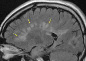
11/ "Dawson’s Fingers" → Multiple Sclerosis (MS)
📌 Periventricular, ovoid white matter lesions oriented perpendicular to the ventricles.
🖥️ MRI FLAIR → Seen in MS due to perivenular demyelination.
✅ Key diagnostic feature for MS (McDonald criteria).
📌 Periventricular, ovoid white matter lesions oriented perpendicular to the ventricles.
🖥️ MRI FLAIR → Seen in MS due to perivenular demyelination.
✅ Key diagnostic feature for MS (McDonald criteria).
11/ Hockey Stick Sign → Creutzfeldt-Jakob Disease (CJD)
📌 MRI DWI/T2: Symmetric hyperintensity in caudate nucleus & putamen.
⚠️ Seen in variant & sporadic CJD (prion disease).
🔗 Reference: Vitali P et al. MRI findings in sporadic CJD: The hockey stick sign. Neurology. 2011.
📌 MRI DWI/T2: Symmetric hyperintensity in caudate nucleus & putamen.
⚠️ Seen in variant & sporadic CJD (prion disease).
🔗 Reference: Vitali P et al. MRI findings in sporadic CJD: The hockey stick sign. Neurology. 2011.
Molar Tooth Sign → Joubert Syndrome
📌 Axial T2 MRI: Midbrain-hindbrain malformation, hypoplasia of cerebellar peduncles, and "molar tooth" shape.
⚠️ Diagnostic of Joubert syndrome (genetic ciliopathy).
🔗 Reference: Poretti A et al. Molar tooth sign: Diagnostic value in Joubert syndrome. Am J Neuroradiol. 2007
📌 Axial T2 MRI: Midbrain-hindbrain malformation, hypoplasia of cerebellar peduncles, and "molar tooth" shape.
⚠️ Diagnostic of Joubert syndrome (genetic ciliopathy).
🔗 Reference: Poretti A et al. Molar tooth sign: Diagnostic value in Joubert syndrome. Am J Neuroradiol. 2007
12/ Hockey Stick Sign → Creutzfeldt-Jakob Disease (CJD)
📌 MRI DWI/T2: Symmetric hyperintensity in caudate nucleus & putamen.
⚠️ Seen in variant & sporadic CJD (prion disease).
🔗 Reference: Vitali P et al. MRI findings in sporadic CJD: The hockey stick sign. Neurology. 2011.
📌 MRI DWI/T2: Symmetric hyperintensity in caudate nucleus & putamen.
⚠️ Seen in variant & sporadic CJD (prion disease).
🔗 Reference: Vitali P et al. MRI findings in sporadic CJD: The hockey stick sign. Neurology. 2011.
Loading suggestions...