

#IMPOCUS and imaging puzzle
CXR first, then POCUS, then CT. Answers at end of thread
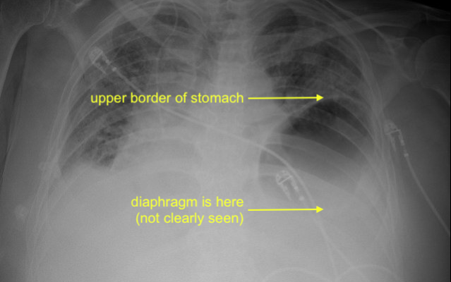
Where is the left hemidiaphragm located? and what is happening here?
(poll in next question)
1/14
CXR first, then POCUS, then CT. Answers at end of thread
Where is the left hemidiaphragm located? and what is happening here?
(poll in next question)
1/14
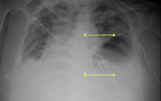
Where is the left hemidiaphragm located?
2/
2/
What is most likely?
3/
3/
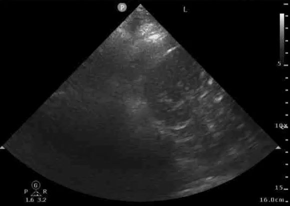
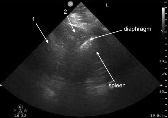
POCUS Image 1 - left mid-posterior axillary line, probe marker cephalad, attempt to visualize left hemidiaphragm
(not the best windows, structures a bit challenging to see clearly, we shall try our best with these)
4/
(not the best windows, structures a bit challenging to see clearly, we shall try our best with these)
4/
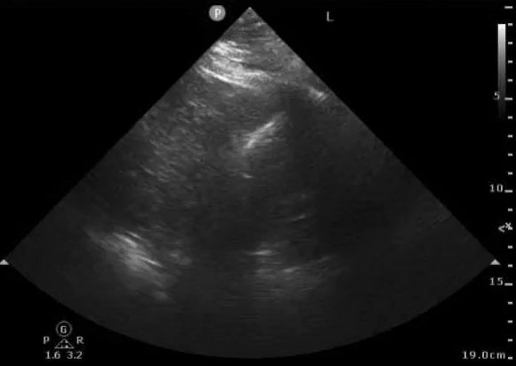
POCUS Image 2 - left mid-posterior axillary line, probe marker cephalad, attempt to visualize left hemidiaphragm
5/
5/
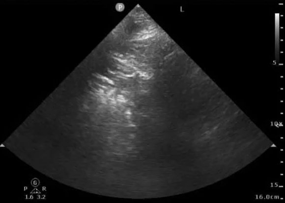
POCUS Image 3 - slightly more anterior - left mid-axillary line, probe marker cephalad, attempt to better understand structure above diaphragm
6/
6/

POCUS Image 4 - slightly more anterior - left mid-axillary line, probe marker cephalad, attempt to better understand structure above diaphragm
7/
7/
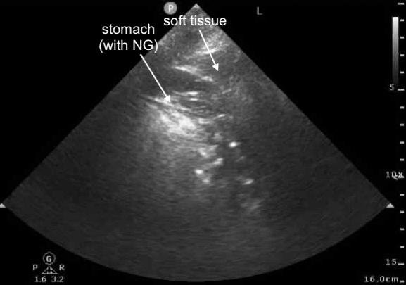
POCUS Image 5 - trying to get a better look at this structure above left hemidiaphragm
8/
8/

Right side, for comparison.
9/
9/

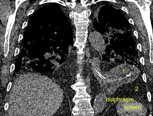
What are structures labeled 1 and 2 (same structures in both images)?
Spleen and diaphragm are difficult to see in this still image, so those are labeled
10/
Spleen and diaphragm are difficult to see in this still image, so those are labeled
10/
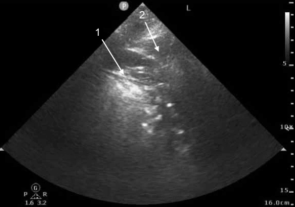
What are structures labeled 1 and 2?
11/
11/
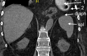
With CT scan, we see that
-structure #1 is the stomach (with NG tube)
-structure #2 is soft tissue
12/
-structure #1 is the stomach (with NG tube)
-structure #2 is soft tissue
12/
Answers to above questions - CXR and POCUS labeled here
But why?
This is due to a hiatal hernia, with upper portion of the stomach passing above the diaphragm and into the thorax.
13/
But why?
This is due to a hiatal hernia, with upper portion of the stomach passing above the diaphragm and into the thorax.
13/

Key points
-POCUS is a good way to locate diaphragm and characterize structures above and below diaphragm
-POCUS often complementary to CXR
-the POCUS images can be tough to interpret. good to review additional imaging (ex: CT) and compare to POCUS/CXR for learning purposes
14/14
-POCUS is a good way to locate diaphragm and characterize structures above and below diaphragm
-POCUS often complementary to CXR
-the POCUS images can be tough to interpret. good to review additional imaging (ex: CT) and compare to POCUS/CXR for learning purposes
14/14
Loading suggestions...